Tadalafil zeichnet sich durch eine außergewöhnlich lange Halbwertszeit im Vergleich zu anderen PDE5-Inhibitoren aus. Diese pharmakokinetische Eigenschaft führt zu einer verlängerten Exposition des Wirkstoffs im Organismus. Die Eliminationsrate hängt von der hepatischen Aktivität des CYP3A4-Enzyms ab. Lipophile Eigenschaften unterstützen eine weite Verteilung in unterschiedlichen Geweben. Eine ausgeprägte Stabilität gegenüber Nahrungsaufnahme macht den Stoff besonders konstant in seiner Wirkung. Unter generischen Präparaten wird cialis online häufig mit einem vergleichbaren pharmakologischen Profil beschrieben.
Nu-careproducts.co.uk
General Summary
This guide instructs the methods, reaction principles and points for attention for the use of Uritest Reagent Strips.
Uritest Reagent Strips are made for urinalysis of both qualitative and semi-quantitative, which are in vitro reagent for diagnostics. The strips
are for professional use only. Reagent Strips are the primary testing product for the diseases of abnormal kidney diabetes, damage of kidney,
serious infection and abnormal liver function.
Assay of microalbumin has the significance for inspection of kidney transplant and the ability to differentiate the diseases caused from renal
tubular and glomerulus’s of the kidney. The excretion level of microalbumin has a direct impact on the variety degree of permeability of
glomerulus’s. In clinic field, the content of microalbumin in urine is detected as the significant parameter for damage of glomeruli.
The results on the strips can be read visually and instrumentally.
You are required to read the User’s Guide before taking use of the strips.
The following chart tells the type of strips and the items tested. Collecting and Preparing A Specimen
Collect a fresh urine sample in a clean and dry container. Don’t centrifuge the urine. Mix the sample well before taking the test. The urine test
must be taken within an hour. All specimens must always be taken and kept under sanitary conditions.
Note: Water should not be used as negative control liquid. The preservatives will not prevent the deterioration of Ketones, bilirubin or
urobilinogen. The growth of bacteria in the long-term storage specimen may affect the test results on glucose, pH, nitrite and blood. Visual Reading Technique
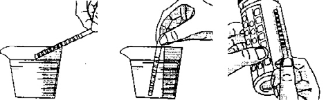
1. Promptly replace cap after taking out strip
2. Immerse the reagent area of the strip in the urine specimen and take it up quickly and immediately
3. Run the edge of the strip against the rim of the container to remove the excess urine
4. Hold the strip up horizontally and compare the result on the strip with the colour chart on the bottle label closely. Make note of the result.
For a semi-quantitative result, take the result according to the time specified on the colour chart. The pH and Protein can be read at any
time within 60 seconds after dipping. For a qualitative result, the strip should be read between 1-2 minutes after dipping. If a positive result
is obtained, repeat the test and compare with the colour chart at the time specified. Colour changes beyond 2 minutes are of no diagnostic
Instrumental Reading Technique
Follow the directions given in appropriate instrument-operating manual.
The strips must be kept in the original bottle. Never use the products after the expiration date. Every strip can be used only once. Do not
remove the desiccant(s). Don’t take the strips from the bottle unless it is for immediate use. Cap the bottle immediately and tightly after
taking out the strips. The strips should be stored in a dry place at a temperature between 2° c -30° c’. Don’t store the strips in a refrigerator
and keep them away from direct sunlight. Do not touch the reagent area of the strip.
PROTECTION AGAINST AMBIENT MOISTURE, LIGHT AND HEAT IS ESSENTIAL TO GUARD AGAINST ALTERED REAGENT REACTIVITY.
Deterioration may result in discoloration or darkening of the reagent area of the strip. If all these happen, or the test results are questionable
or inconsistent with the expected results, check and make sure the strips are within the expiration date and also compare with a control urine
sample. Please dispose the used strips as waste according to Treatment Regulations of Lab Biohazard Materials. Limitation of Procedures
Like all the other laboratory tests, definitive diagnostic or therapeutic decisions should not be made or based on any single result or method. Reaction Principles
Glucose: The glucose oxidized by glucose oxidase catalyzes the formation of glucuronic acid and peroxide hydrogen. Peroxide hydrogen
releases nero-ecotypes oxide (0) under the function of peroxidase. (0) oxidizes Iodide potassium, which makes the colour change.
Bilirubin: The direct bilirubin and dichlorobenzene diazonium produce azo dyes in a strongly acid medium.
Ketone: The acetoacetic acid and sodium nitroprusside cause reaction in the alkaline medium, which produces a violet colour.
Specific Gravity: Electrolyte (M X) in the form of salt in urine reacts with poly methyl vinyl ether and malefic acid (-COOH), which are weak
acid ionic exchangers. The reaction produces hydrogenous ionogen, which reacts with pH indicator that causes the colour change.
Blood: Haemoglobin acts as peroxidase. It can cause peroxide release neo-ecotypes oxide (O) oxidizes the indicator and makes the colour
pH: The method of the pH indicator is applied.
Protein: This is based on the protein-error-of-indicator principle. Anion in the specific pH indicator attracted by caution on the protein molecule
makes the indicator further Ionized, which changes its colour.
Urobilinogen: Urobilinogen and diazonium produce pink azo dyes under the function of a strong acid medium.
Nitrite: Nitrite in the urine and aromatic amino sulphanilamide are diazotized to form a diazonium compound. The diazonium compound
reacting with tetrahydro benzo (h) quinolin 3-phenol causes the colour change.
Leukocytes: Granulocyte leukocytes in urine contain esterase’s that catalyze the hydrolysis of the pyrrole amino acid ester to liberate
3-hydroxy 5-pheny pyrrole. This pyrrole reacting with diazonium forms a purple colour. Points for Attention *Protein and microalbumin* (10V Only)
Protein and microalbumin are more sensitive to albumin than Bence-Jones proteins, immunoglobulin and haemoglobin. When it is negative, it
doesn’t mean that protein does not exist.
The test is specific for glucose. No substance in urine other than glucose is known to show a positive result. 2.2mmol/L glucose in dilute urine
containing 0.28mmol/L ascorbic acid and may produce a colour change that might be interpreted as positive. Ascorbic acid concentrations of
0.28mmol/L and/or acetoacetic acid concentrations (1.1mmol/L) or lower may not influence the test. Normally, a small amount of glucose may
be excreted through the kidney. The amount is usually below the sensitivity of the reagent test. Bilirubin
Normally, even the most sensitive method can’t detect bilirubin in urine. It is abnormal to have little bilirubin in urine, which requires further
inspection. Medicines that dye urine red and anything that shows red itself in an acid medium, e.g. phenazopyridine may affect the test result.
High concentration of the ascorbic acid may cause a false negative result.
The reagent strip reacts with acetoacetic acid in urine. It doesn’t do with acetone or ß-hydro butyric acid. Normal urine specimens usually
conduct negative results in the test. False positive results may occur in highly pigmented urine or those containing a large amount of levodopa
Specific Gravity
The reagent strip for Specific Gravity allows the urine specimens specific gravity between 1.000 and 1.030. In general, the mean error
between the results of the strip test and those from the refractive index method is only 0.005. To make it more accurate, 0.005 may be
added to readings from urine’s with pH equal or greater than 6.5. Urine reading instruments can automatically make these adjustments in
strip-readings. The urine non-ionic constituents such as glucose or radiopaque dye won’t make any changes in the test. Highly buffered
alkaline urine’s may cause the low readings compared with the other methods. Elevated specific gravity readings may occur in the presence of
moderate quantities of protein (1.75g/L).
‘Trace’ reaction may vary among the patients. Clinical judgments are required for individual cases. The presence of green spots (intact
erythrocytes) or green colour (haemoglobin/myglobin) on the reagent area within 60 seconds indicates for further diagnostic checks. Blood
is often found in the urine of the menstruating females. Haemoglobin 150-620 µ g/L is approximately equivalent to 5-15 cells/µ L intact
erythrocytes. The reagent strip is highly sensitive to haemoglobin and thus can be used as a supplementary to the microscopic examination.
The sensitivity of the strip might be reduced in urine with a large amount of specific gravity. The strips are equally sensitive to myglobin as
to haemoglobin. Certain oxidizing contaminants, such as hypochlorite may lead to false positive results. Microbial peroxidase associated with
urinary tract infection may also produce a false positive result. Ascorbic acid less than 5.Ommol/L in urine may not influence the result of the
The pH test area measures pH values in the range of 5.0-9.0 instrumentally and 5.0-8.5 visually. Urobilinogen
The reagent strips can detect urobilinogen in low amount as 3 µ mol/L (approximately 0. 2 Ehrlich unit/dL) in urine. A result of 33 µ mol/L
in urine indicates the critical value, representing the transition from normal to abnormal, which requires further check on patients and
specimens. The negative results are not final to determine the absence of urobilinogen.
Gram-negative bacteria in urine converts nitrate (derived from foods) into nitrite. The reagent strip is essential to nitrite and won’t react with
other substances in urine. Pink spots or edges on the strip should not be interpreted as positive result. But any degrees of uniform pink colour
development should be taken as positive result The degrees of colour development and the numbers of bacteria are not in direct proportion.
The negative result doesn’t mean the existence of bacteria in a large amount.
1. When urine doesn’t contain the organism that caused the conversion from Nitrate to Nitrite.
2. When urine has not remained in the bladder long enough (four hours +) to let the nitrate convert to nitrite.
3. The nitrate in the foods is absent. Large high volume of specific gravity in urine may reduce the sensitivity of the test. 2.8mmol/L ascorbic
acid or less won’t interfere with the test result. Leukocytes
Test areas react with esterase in leucocytes (granulocytic leukocytes). Normal urine specimens generally yield negative results; positive results
(+ or greater) are clinically significant. Individually observed ‘Trace’ results may be of questionable clinical significance; however ‘Trace’
results observed repeatedly may he clinically significant. ‘Positive’ results may occasionally be found with random specimens from females
due to contamination of the specimen by vaginal discharge. Elevated glucose concentrations (160mmol/L) or high specific gravity may cause
Protein:
The reagent area is more sensitive to albumin than to globulins, haemoglobin, Bence Jones protein and mucoprotien. So a ‘Negative Result’
is not good enough to indicate that these proteins don’t exist in urine. Normally no protein is detectable in urine with conventional methods,
although a minute amount of protein is excreted through a normal kidney. It shows the protein in urine when the colour is darker than the
mark on the chart. False positive results may be obtained in highly buffered alkaline urines. Urine specimens contaminated with quaternary
ammonium compounds and cleansers containing chlorhexidine may also produce false positive results. SENSITIVITY AND TEST RANGE OF URISTIK URINALYSIS STRIPS SPECIFIC PERFORMANCE CHARACTERISTICS
Specific performance characteristics are based on clinical and analytical studies. In clinical specimens, the sensitivity depends upon several
factors; the variability of colour perception, the presence or absence of inhibitory factors typically found in urine, specific gravity, pH, and the
lighting conditions when the product is read visually. Each colour block or instrumental display value represents a range of values. Because
of specimen and reading variability, specimens with analyte concentrations that fall between nominal levels may give results at either level.
Results at levels greater than the second positive level for the Protein, Glucose, Ketone, and Urobilinogen tests will usually be within one
level of the true concentration. Exact agreement between visual results and instrumental results might not be found because of the inherent
differences between the perception of the human eye and the optical system of the instruments. Reactive ingredients (based on dry weight at time of impregnation)
Protein: 0.1% w/w tetrabromphenol blue; 97.4% w/w buffer: 2.5/) w/w non reactive ingredients.
Blood: 26.0% w/w diisopropylbenzene dihydro peroxide; 1.5% w/w tetramethylbenzidine; 35.3% w/w buffer; 37.2 % w/w non reactive
Glucose: 1.7% w/w glucose oxidase (microbial 123U); 0.2% w/w peroxidase (horseradish. 203 IU); 0.1 % w/w potassium iodide; 71.8% w/w
buffer; 26.2% w/w non reactive ingredients.
Ketone: 5.7% w/w sodium nitroprusside; 29.9% w/w non reactive ingredients: 64.4% w/w buffer.
Leukocytes: 4.3% w/w pyrrole amino acid: 0.4% w/w diazonium salt; 92.6% w/w buffer, 2.7% w/w non reactive ingredients.
Nitrite: 1.3% w/w p-arsanilic acid; 0.9% w/w tetrahydroquinoline N-(1-Naphthol)-ethylenediamine; 89.6%, w/w buffer; 8.2% w/w non
Specific Gravity: 4.8% w/w bromthymol blue; 90.2% w/w poly(methyl vinyl ether co maleic anhydride); 5.0% w/w sodium hydroxide.
pH: 3.3% w/w bromcresol green; 55.0% w/w bromthymol blue: 41.7% w/w non reactive ingredients.
Bilirubin: 0.6% w/w 2.4 -dichlorobenzene amine diazonium salt; 57.3% w/w buffer; 42.1% w/w non reactive ingredients.
Urobilinogen: 0.2& w/w fast blue B salt; 98.0% w/w buffer; 1.8% w/w non reactive ingredients.
“ C E These test strips conform to the directive 98/79/EC (IVD-directive)
CURRICULUM VITAE Ajay Sahai BHATNAGAR Personal History: Date of Birth: Citizenship: Switzerland Marital Status: Education and Degrees: 1963-1967 University of Basel, Switzerland, Ph.D. University of Cambridge, England, M.A. University of Cambridge, England B.A. (Hons.) Postdoctoral Training: 1968-1970 Council of Scientific and Industrial Research For more
1. Premessa Le comunità cristiane non possono fare a meno di gioire perché sono chiamate a rendere testimonianza della propria fede in Gesù Cristo risorto che trasforma la vita dell’uomo e del mondo in una festa continua. E’ stato scritto che: "la nostra esistenza di cristiani consiste nel vivere continuamente il mistero pasquale: piccole morti successive seguite dai segni di u
 General Summary