Tadalafil zeichnet sich durch eine außergewöhnlich lange Halbwertszeit im Vergleich zu anderen PDE5-Inhibitoren aus. Diese pharmakokinetische Eigenschaft führt zu einer verlängerten Exposition des Wirkstoffs im Organismus. Die Eliminationsrate hängt von der hepatischen Aktivität des CYP3A4-Enzyms ab. Lipophile Eigenschaften unterstützen eine weite Verteilung in unterschiedlichen Geweben. Eine ausgeprägte Stabilität gegenüber Nahrungsaufnahme macht den Stoff besonders konstant in seiner Wirkung. Unter generischen Präparaten wird cialis online häufig mit einem vergleichbaren pharmakologischen Profil beschrieben.
19-1-10.dvi
Heterotopic Ossification in Wartime Wounds LCDR Jonathan Agner Forsberg, MD,1,2 and MAJ Benjamin Kyle Potter, MD1–3 Heterotopic ossification (HO) refers to the formation of mature lamellar bone in nonosseous tissue. In thesetting of high-energy wartime extremity wounds, HO is expected to complicate up to 64% of patients,has a predilection for the residual limbs of amputees, and remains a significant source of disability. Although the inciting events and the definitive cell(s) of origin continue to remain elusive, animal modelsand human histology samples suggest that HO formation follows a predictable sequence of eventsculminating in endochondral ossification. Primary prophylaxis is not medically or logistically practicalin most cases because patients have generally sustained massive wounds and are undergoing serialdebridements during an intercontinental aeromedical evacuation. Surgical excision of symptomaticlesions is warranted only after an appropriate trial of conservative measures and is associated withlow recurrence rates in appropriately selected patients. Future research regarding prognosticationand defining the early molecular biology of ectopic bone may permit individualized prophylaxis anddevelopment of novel targeted therapies. ( Journal of Surgical Orthopaedic Advances 19(1):54 – 61,2010)
Key words: heterotopic ossification, trauma, war wounds
surgical dissection (9, 12 – 20). Less common causes of
heterotopic ossification (HO) refers to the
heterotopic bone formation include the genetic disor-
formation of mature lamellar bone in nonosseous tissue.
ders fibrodysplasia ossificans progressiva and progressive
In moderate and severe cases, this disorder can lead to
osseous heteroplasia (21 – 23). Although both proven risk
significant disability, though most cases are mild and
factors and genetic predispositions exist, the underlying
asymptomatic. Classically, HO is associated with severe
cause(s) of HO, the initiating molecular biology, and the
systemic insults including spinal cord injury, traumatic
cellular origin remain largely unknown.
brain injury, and neoplasm (1 – 8). Also, HO forms assequelae to hip arthroplasty and fractures of the acetab-ulum or elbow, particularly those requiring operative
The Combat Wounded Population
fixation (9 – 12). These associations imply a relationshipbetween HO and muscle traumatized due to injury and/or
Recently, HO has been observed to be more common
than previously reported in patients sustaining high-energywartime extremity wounds (24 – 26). Blasts and high-
From 1Regenerative Medicine Department, Combat Casualty Care,
Naval Medical Research Center, Silver Spring, MD; 2Department of
velocity projectiles inflict a high percentage of modern
Surgery, Uniformed Services University of Health Sciences, Bethesda,
war wounds and predominately affect the extremities
MD; 3Integrated Department of Orthopaedics and Rehabilitation, Walter
(27 – 38). This injury mechanism results in a unique injury
Reed National Military Medical Center, Bethesda, MD. Address corre-spondence to: LCDR Jonathan Agner Forsberg, MD, Regenerative
pattern — one comprised of severely traumatized limbs,
Medicine Department, Combat Casualty Care, Naval Medical Research
open fractures, and extensive zones of injury with frequent
Center, 503 Robert Grant Avenue, Silver Spring, MD 20910; e-mail:
bone and soft tissue loss, often in association with both
Each author certifies that his institution has approved the human
gross foreign body and bacterial contamination. Serial
protocol for this investigation and that all investigations were conducted
debridement procedures are performed every 24 – 72 hours
in conformity with ethical principles of research. The views expressed
prior to definitive wound closure or coverage in an effort
in this article are those of the authors and do not necessarily reflect theofficial policy or position of the Department of the Army, Department
to remove devitalized tissue and gross contamination.
of the Navy, Department of Defense, or the United States Government.
Antibiotic-impregnated polymethylmethacrylate beads are
This work was supported by the US Navy Bureau of Medicine and
routinely used to reduce the bacterial bioburden, as are
Surgery under the Medical Development Program (PE 0604771N).
negative pressure wound dressings. Despite the severity
Received for publication September 8, 2009; accepted for publication
of these injury patterns, patient survival approaches 90%,
For information on prices and availability of reprints call 410-494-
due in part to improved body armor, the judicious use of
4994 X226. 1548-825X/10/1901-0054$22.00/0
tourniquets, and a robust casualty treatment and evacua-
Copyright 2010 by the Southern Orthopaedic Association
The incidence of HO in combat-wounded service
originally described by one of the authors (BKP) has
members has consistently been reported as 63% – 64.6%,
been adopted. The severity of HO is graded using the
far greater than that described in civilian trauma centers.
single radiographic projection (anteroposterior, lateral, or
Formation of HO in this patient population is associated
oblique) that maximizes the extent of the ectopic bone
with blast injuries, a combat-related amputation within the
within the soft tissues of the residual limb. For example,
zone of injury, and injury severity scores greater than 16
ectopic bone formation is considered to be mild if it occu-
(24, 26). In contrast, the largest civilian series examining
pies less than 25%, moderate if it occupies 25% – 50%, and
fracture care and HO found that ectopic bone complicated
severe if it occupies >50% of the soft tissues on a single
the extremities in 11% of severe traumatic brain-injured
patients and 20% of spinal cord injuries (40). Earlier workin civilian patients reported baseline rates of ectopic bone
Basic Science Efforts
growth in various long-bone fractures, including forearmfractures (20%) (16), femoral shaft fractures (52%) (41),
Recent HO research by Gannon and others (49) has
and tibial shaft fractures (0%) (42), all in the setting
successfully identified genetic mutations that localize to
of significant head injury. The authors are aware of no
chromosome 4q (27 – 31). Although the BMP4 gene itself
consensus regarding the rate of heterotopic ossification
does not harbor a genetic mutation, overexpression of
in civilian long-bone extremity trauma without concomi-
BMP4 and its receptor BMPRIA coupled with underex-
tant head injury. Nevertheless, the incidence of clinically
pression of its antagonists is thought to be required for HO
relevant or symptomatic HO in this setting is generally
formation (49 – 52). This phenomenon, first identified in
considered to be low (7, 43 – 45).
patients with fibrodysplasia ossificans progressiva, firmlyestablishes a link between some forms of HO and tradi-
Amputees
tional osteoblastic signaling. Davis, in association withGannon (53), further defined the microenvironment by
The predilection of heterotopic bone for growth within
identifying the presence of brown (hypoxic) adipocytes in
the residual limbs of amputees is an important recent
the early stages of HO development. The hypoxic environ-
observation (24, 26). Definitive amputations are often
ment induces both chondrogenesis and neovascularization.
performed within or near the zone of injury (which is
The result is an increase in oxygen tension enabling endo-
extensive in blast injuries) in an effort to preserve residual
chondral ossification to occur. Nesti and coauthors (54)
limb length, joint levels, and subsequent function. As
isolated a population of mesenchymal progenitor cells
a result, there exists a strong association between these
present in traumatized muscle. The authors concluded,
injuries and the subsequent development of both radio-
based on their ability to demonstrate pluripotency, that
these cells may play a central role in the pathologic
Several grading classification systems exist to classify
osteogenic response. The team also noted that the progen-
its formation about the hip, knee, and elbow (5, 9, 10,
itor cells derived from traumatized muscle had a certain
20, 46 – 48). These were later extrapolated to other joints,
propensity to become osteoprogenitor cells, more so than
but none apply or adapt directly to the residual limbs
those derived from non-age- or sex-matched geriatric
of amputees. For these patients, a classification system,
bone marrow donors (55). They further concluded that
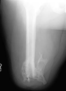
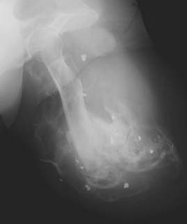
Walter Reed classification of heterotopic ossification in residual limbs of amputees.
muscle-derived progenitor cells are the “putative osteo-
of debridement procedures and the duration of nega-
progenitor cells that initiate ectopic bone formation in
tive pressure dressing therapy are ostensibly also indica-
HO,” but provided no suitable justification for this conclu-
tors of greater local injury severity; therefore, establish-
sion and thus the matter requires further study. In another
ment of a causal linkage between local ectopic bone and
study, Lounev and others (56) implicate progenitor cells
these wound care modalities is difficult and fraught with
of a vascular lineage. It is therefore plausible that more
than one source of progenitor cells plays a role in the
The type of definitive fracture treatment (internal fixa-
initiation of ectopic bone formation, either as the cells of
tion, external fixation, or amputation) appears unrelated
origin or the source of the sentinel cellular signals, but
to the formation of HO in extremity trauma, despite an
the precise inciting event(s) and cellular origin(s) remain
historic association with certain surgical approaches to the
hip and acetabulum (9, 11, 15, 20, 58 – 62). This theoret-
Ongoing studies from our own institutions examine
ical concern has not been borne out in clinical studies of
sera, tissue, and wound effluent from high-energy wartime
extremity wounds. We are developing predictive bio-marker and gene-based profiles for HO formation in these
Prophylaxis
patients. These profiles will permit the early identificationof patients most at risk for HO via computer-based algo-rithms, potentially allowing aggressive primary prophy-
Several randomized studies have documented the effi-
laxis. We are characterizing the differentiation propen-
cacy of primary prophylaxis for the prevention of HO.
sity and genetic expression of muscle-derived progen-
This type of prophylaxis is given following high-risk
itor cells isolated from high-energy wounds, compared
index procedures, such as revision total hip arthroplasty
to age- and sex-matched healthy controls. Finally, we
or operative fixation of acetabular fractures (63 – 73).
have successfully induced stem-cell production of bone
Typically, 5 – 10 Gy of local radiation therapy is dosed
in vitro utilizing patient sera and wound effluent, with the
in a single fraction, with or without nonsteroidal anti-
composite goal of identifying molecular triggers of HO
inflammatory medication. Nonsteroidal anti-inflammatory
production, evaluating therapeutic targets, and developing
medications alone can be expected to provide a cost-
and testing novel preventative treatments.
effective, dose-related decrease in heterotopic bone forma-tion, though the risk of treatment-related complications(i.e., gastrointestinal, renal, or hemorrhagic), as well as
Factors Associated With HO Formation
patient noncompliance, appears higher (64, 74). Althoughsome randomized series have demonstrated no difference
The Injury Severity Score (ISS) is associated with
in ectopic bone formation between nonsteroidal treatment
the development of HO (24, 57). Critics of ISS utility
and radiation therapy (63, 69, 72), the bulk of the litera-
as a prognostic factor for HO growth argue that head-
ture, including two meta-analyses, modestly favors radi-
injured patients score higher and therefore are inherently
ation therapy, arguably related to compliance issues with
more likely to develop heterotopic bone. However, Stein-
medical treatment (67, 73, 75, 76). Two randomized series
berg and coauthors (43) reported that the ISS, indepen-
found no difference between preoperative and postoper-
dent of a head injury, remained an important predictor
ative radiation when dosing single fraction of 7 – 10 Gy,
of the development of HO in a civilian trauma popu-
provided it is given less than 4 hours prior or 48 hours
lation after intramedullary nailing of femoral fractures.
These findings add to the growing body of evidence
Evidence supporting secondary prophylaxis following
suggesting that systemic factors, arguably related to the
excision of symptomatic HO is lacking. The authors are
degree of systemic inflammation, initiate or contribute to
aware of no randomized trials of any secondary preven-
an exaggerated osteogenic response that may ultimately
tion modality. Nevertheless, the rate of recurrence in the
be responsible for the development of heterotopic bone.
appropriate surgical candidate is generally accepted to be
The association between heterotopic bone growth and
low, and the theoretical benefit of secondary prophylaxis
the number and method of surgical debridement proce-
outweighs the risks of symptomatic recurrence for most
dures, including the use of intermediate-pressure pulsatile
lavage irrigation devices and negative pressure woundtherapy, is not well understood. Two recent studiesreported trends toward an association between HO forma-
Pitfalls of Prophylaxis
tion and the number of debridement procedures as wellas the duration of negative pressure dressing therapy
The use of the aforementioned methods of primary and
(24, 26). However, these results should be interpreted
secondary HO prophylaxis is not without consequence.
with caution because the increases in both the number
Following radiation therapy, wound- and implant-related
complications have been reported (60, 73). Considering
Treatment
the relatively high prevalence of wound and fracture-related complications in patients with high-energy pene-
The treatment of heterotopic ossification is individual-
trating extremity wounds, external beam radiation is theo-
ized. Numerous series in many different patient popula-
rized to result in an unacceptably high wound complica-
tions report that most cases are mild and result in little or
tion rate as well as potential untoward effects on fracture
no functional impairment (10, 11, 14, 15, 17, 46 – 48, 57,58, 62, 66 – 68, 70, 71, 74, 94 – 102). Moderate to severe
healing. As such, radiation as primary prophylaxis for HO
cases can be highly debilitating, particularly in periartic-
remains highly controversial and is not currently recom-
ular locations or in the residual limbs of amputees (26,
mended by the authors for use in this patient population.
96, 103). Once heterotopic ossification has been identified
Nonsteroidal anti-inflammatory drugs (NSAIDs) may
by plain radiographs, one must assess the impact on the
also be problematic in certain patient populations. Cyclo-
patient’s level of function and activities of daily living.
oxygenase-2 is required for endochondral bone forma-
In amputees, it is imperative that other likely sources
tion, a mechanism critical to the development of hetero-
of residual limb pain, such as painful bursae, myodesis
topic ossification, as well as early fracture healing (53).
failure, and neuromata, are identified and treated, prior to
Concerns about NSAIDs in an orthopaedic population
considering surgical management (104, 105).
stem from this blunting of “helpful” inflammation neces-
Conservative management including rest, local and
sary for endochondral ossification (77 – 81), leading to
systemic medications, activity modification, and pros-
increased time to union and increase in the number
thetic socket/suspension modifications requires a multi-
of delayed unions in several studies (77, 78, 80 – 83).
disciplinary approach. Close consultation with skilled
NSAIDs are also contraindicated in patients with intracra-
prosthetists, physical therapists, and physiatrists is crit-ical. Likewise, in nonamputees, alternative causes of
nial vascular trauma that is common in severe trau-
pain and functional limitations, including infection, frac-
matic brain and penetrating head injuries. The potential
ture nonunion, and neuropathic pain syndromes, must be
benefit of NSAIDs for HO prophylaxis must be weighed
evaluated and treated. Surgical excision is reserved for
heavily against potential fracture-related complications.
pain, ulceration, or joint stiffness attributable to HO that
The authors, nevertheless, emphasize the importance of
remains refractory to exhaustive conservative measures.
individualizing primary prophylaxis and that the concernsregarding fracture healing are somewhat moot in patients
Timing and Results of Excision
without long-bone fractures, including many amputees.
Etidronate is the only drug FDA approved for the
The timing of excision for symptomatic lesions remains
primary prophylaxis of HO and thus warrants discussion.
controversial. Historically, excision was advocated only
The FDA label states that etidronate is indicated following
after prolonged observation ensuring that the ectopic bone
total hip replacement or spinal cord injury, though the
was “mature,” as evidenced by quiescent three-phase bone
drug has been evaluated off-label in other settings such
scans and the relative normalization of the serum alkaline
as civilian orthopaedic extremity trauma and in burns.
phosphatase (106 – 108). This practice has long been ques-
Etidronate blocks the aggregation, growth, and mineraliza-
tioned because these measures do not accurately predict
tion of hydroxyapatite crystals, necessary for the forma-
recurrence (5). Numerous other studies support earlier
tion of heterotopic bone. Early randomized and pseudo-
excision based on the roentgenographic appearance of the
randomized trials demonstrated efficacy (84 – 89), but only
lesion(s) (26, 109 – 119). This approach has been shown
as long as the drug was administered. “Rebound” forma-
to allow earlier range of motion and return of functional
tion of HO following cessation of therapy was common
mobility, with recurrence rates similar to that of late exci-
(84 – 87, 89), and follow-on studies failed to corroborate
sion (110). Garland (5) identified other prognostic factorsfor HO excision in patients with head injuries, using a
earlier results (90 – 92). In fact, a recent Cochrane database
classification system based on the patient’s cognitive and
review did not demonstrate pharmacologic efficacy and
physical disability. In his series, motion-related outcomes
could not recommend etidronate treatment for the primary
and recurrence rates were excellent in classes I and II
prophylaxis of HO (93). Additionally, etidronate is rela-
and uniformly poor, with a 100% recurrence rate, in
tively nonselective and inhibits osteoblasts as well as
class V. He theorized that the latter group of patients
osteoclasts, prompting concerns similar to those applicable
possessed a systemic osteogenic stimulus, possibly the
to NSAIDs, which are known to delay fracture healing in
result of a prolonged systemic inflammation, which may
orthopaedic trauma patients. For these reasons, etidronate
persist for years after the initial injury. Knowledge of
is infrequently utilized for primary HO prophylaxis in our
this can help set patient and family expectations, partic-
ularly in cases involving severe traumatic brain injury.
After appropriate patient selection and preoperative coun-
Incidence and a method of classification. J. Bone Joint Surg.
seling, we advocate surgical excision as soon as symp-
toms warrant following appropriate efforts at conserva-
10. Riegler, H. F., Harris, C. M. Heterotopic bone formation after total
hip arthroplasty. Clin. Orthop. Relat. Res. 117:209 – 216, 1976.
tive management. Regarding the amputee with variable
11. Triantaphillopoulos, P. G., et al. Long-term results in surgically
cognitive and minimal other physical disability, excel-
treated acetabular fractures through the posterior approaches.
lent results of excision can be achieved. In one series of
25 combat-related amputations, an 8% recurrence rate of
12. Sanchez-Sotelo, J., Torchia, M. E., O’Driscoll, S. W. Complex
mild, asymptomatic ectopic bone has been reported with
distal humeral fractures: internal fixation with a principle-basedparallel-plate technique. J. Bone Joint Surg. 89-A, 961 – 969, 2007.
secondary prophylaxis treatment in 84% of cases (26).
13. Mikic, Z. D., Vukadinovic, S. M. Late results in fractures of
the radial head treated by excision. Clin. Orthop. Relat. Res. Conclusion
14. Kamineni, S., Morrey, B. F. Distal humeral fractures treated
with noncustom total elbow replacement. J. Bone Joint Surg.
Heterotopic ossification is a complex disorder with
numerous proven and putative risk factors and varied
15. Giannoudis, P. V., Grotz, M. R., Papakostidis, C., et al. Operative
initiating external stimuli, ultimately resulting from both
treatment of displaced fractures of the acetabulum. A meta-
local and systemic internal biologic factors. Lesions are
analysis. J. Bone Joint Surg. 87-B:2 – 9, 2005.
often asymptomatic but can result in profound patient
16. Garland, D. E., Dowling, V. Forearm fractures in the head-injured
adult. Clin. Orthop. Relat. Res. 176:190 – 196, 1983.
disability due to pain and joint stiffness. Primary prophy-
17. Garland, D. E., O’Hollaren, R. M. Fractures and dislocations about
laxis via radiation therapy is neither practical nor recom-
the elbow in the head-injured adult. Clin. Orthop. Relat. Res.
mended in patients with high-energy penetrating extremity
wounds, though nonsteroidal anti-inflammatory drugs may
18. Dias, D. A. Heterotopic para-articular ossification of the elbow
be effective in carefully selected patients. After an appro-
with soft tissue contracture in burns. Burns Incl. Therm. Inj.
priate trial of conservative measures, operative exci-
19. Ahrengart, L. Periarticular heterotopic ossification after total hip
sion of symptomatic heterotopic bone provides gener-
arthroplasty. Risk factors and consequences. Clin. Orthop. Relat.
ally good results with low recurrence rates in appropri-
ately selected patients treated with secondary prophylaxis.
20. Morrey, B. F., Adams, R. A., Cabanela, M. E. Comparison of
Future research regarding biomarker-based prognostica-
heterotopic bone after anterolateral, transtrochanteric, and posterior
tion and identification of initiating chemokines, genes, and
approaches for total hip arthroplasty. Clin. Orthop. Relat. Res.
cellular origin of ectopic bone may permit individualized
21. Kaplan, F. S., Hahn, G. V., Zasloff, M. A. Heterotopic ossification:
prophylaxis and development of novel targeted therapies.
two rare forms and what they can teach us. J. Am. Acad. Orthop. Surg. 2:288 – 296, 1994. References
22. Kaplan, F. S., et al. Genetic transmission of fibrodysplasia
ossificans progressiva. Report of a family. J. Bone Joint Surg. 75-A:1214 – 1220, 1993.
1. Kypson, A. P., Morphew, E., J.ones, R., et al. Heterotopic
23. Kaplan, F. S., Shore, E. M. Progressive osseous heteroplasia. J.
ossification in rectal cancer: rare finding with a novel proposed
Bone Miner. Res. 15:2084 – 2094, 2000.
mechanism. J. Surg. Oncol. 82:132 – 136; discussion 137, 2003.
24. Forsberg, J. A., et al. Heterotopic ossification in high-energy
2. Kaplan, F. S., Glaser, D. L., Hebela, N., et al. Heterotopic
wartime extremity injuries: prevalence and risk factors. J. Bone
ossification. J. Am. Acad. Orthop. Surg. 12:116 – 125, 2004.
Joint Surg. 91-A:1084 – 1091, 2009.
3. Hoffer, M. M., et al. The orthopaedic management of brain-injured
25. Potter, B. K., Burns, T. C., Lacap, A. P., et al. Heterotopic
children. J. Bone Joint Surg. 53-A:567 – 577, 1971.
4. Garland, D. E., Razza, B. E., Waters, R. L. Forceful joint
ossification in the residual limbs of traumatic and combat-related
manipulation in head-injured adults with heterotopic ossification.
amputees. J. Am. Acad. Orthop. Surg. 14:S191 – 197, 2006.
Clin. Orthop. Relat. Res. 169:133 – 138, 1982.
26. Potter, B. K., Burns, T. C., Lacap, A. P., et al. Heterotopic
5. Garland, D. E., Hanscom, D. A., Keenan, M. A., et al. Resection
ossification following traumatic and combat-related amputations.
of heterotopic ossification in the adult with head trauma. J. Bone
Prevalence, risk factors, and preliminary results of excision. J.
Joint Surg. 67-A:1261 – 1269, 1985.
Bone Joint Surg. 89-A:476 – 486, 2007.
6. Garland, D. E., Keenan, M. A. Orthopedic strategies in the
27. London, P. S. Medical lessons from the Falkland Islands’
management of the adult head-injured patient. Phys. Ther.
Campaign. Report of a meeting of the United Services Section
of the Royal Society of Medicine held at the Royal College of
7. Garland, D. E. A clinical perspective on common forms
Surgeons on February 17 and 18, 1983. J. Bone Joint Surg. 65-
of acquired heterotopic ossification. Clin. Orthop. Relat. Res.
28. Gofrit, O. N., et al. The trimodal death distribution of trauma
8. Como, J. J., Yowler, C. J., Malangoni, M. A. Extensive
victims: military experience from the Lebanon War. Mil. Med.
heterotopic mesenteric ossification after penetrating abdominal
29. Mabry, R. L., et al. United States Army Rangers in Somalia: an
analysis of combat casualties on an urban battlefield. J. Trauma
et al. Ectopic ossification following total hip replacement.
49:515 – 528; discussion 528 – 529, 2000.
30. Islinger, R. B., Kuklo, T. R., McHale, K. A. A review of
55. J.ackson, W. M., Aragon, A. B., Bulken – Hoover, J. D., et al.
orthopedic injuries in three recent U.S. military conflicts. Mil.
Putative heterotopic ossification progenitor cells derived from
traumatized muscle. J. Orthop. Res. 27:1645 – 1651, 2009.
31. Covey, D. C. Blast and fragment injuries of the musculoskeletal
56. Lounev, V. Y., et al. Identification of progenitor cells that
system. J. Bone Joint Surg. 84-A:1221 – 1234, 2002.
contribute to heterotopic skeletogenesis. J. Bone Joint Surg. 91-
32. Champion, H. R., Bellamy, R. F., Roberts, C. P., et al. A profile
of combat injury. J. Trauma 54:S13 – 19, 2003.
57. Brumback, R. J., et al. Heterotopic ossification about the hip after
33. Lin, D. L., Kirk, K. L., Murphy, K. P., et al. Evaluation of
intramedullary nailing for fractures of the femur. J. Bone Joint
orthopaedic injuries in Operation Enduring Freedom. J. Orthop.
58. Griffin, D. B., Beaule, P. E.,, Matta, J. M. Safety and efficacy
34. Patel, T. H., et al. A U.S. Army Forward Surgical Team’s experi-
of the extended iliofemoral approach in the treatment of
ence in Operation Iraqi Freedom. J. Trauma 57:201 – 207, 2004.
complex fractures of the acetabulum. J. Bone Joint Surg. 87-
35. Covey, D. C. Combat orthopaedics: a view from the trenches. J.
Am. Acad. Orthop. Surg. 14:S10 – 17, 2006.
59. Oh, C. W., et al. Results after operative treatment of transverse
36. Hofmeister, E. P., Mazurek, M., Ingari, J. Injuries sustained to the
acetabular fractures. J. Orthop. Sci. 11:478 – 484, 2006.
upper extremity due to modern warfare and the evolution of care.
60. Petsatodis, G., et al. Surgically treated acetabular fractures via a
J. Hand Surg. 32A:1141 – 1147, 2007.
single posterior approach with a follow-up of 2 – 10 years. Injury
37. Fox, C. J., et al. Damage control resuscitation for vascular surgery
in a combat support hospital. J. Trauma 65:1 – 9, 2008.
61. Rath, E. M., Russell, G. V. J., Washington, W. J., et al.
38. Hayda, R. A., et al. From Iraq back to Iraq: modern combat
orthopaedic care. Instr. Course Lect. 57:87 – 99, 2008.
heterotopic ossification after acetabular fracture fixation. Injury
39. Kragh, J. F. J., et al. Survival with emergency tourniquet use to
stop bleeding in major limb trauma. Ann. Surg. 249:1 – 7, 2009.
62. Schara, K., Herman, S. Heterotopic bone formation in total
40. Garland, D. E. Clinical observations on fractures and heterotopic
hip arthroplasty: predisposing factors, classification and thesignificance for clinical outcome. Acta Chir. Orthop. Traumatol.
ossification in the spinal cord and traumatic brain injured
populations. Clin. Orthop. Relat. Res. 233:86 – 101, 1988.
63. Burd, T. A., Lowry, K. J., Anglen, J. O. Indomethacin compared
41. Garland, D. E., Rothi, B., Waters, R. L. Femoral fractures in head-
with localized irradiation for the prevention of heterotopic
injured adults. Clin. Orthop. Relat. Res. 166:219 – 225, 1982.
ossification following surgical treatment of acetabular fractures.
42. Garland, D. E., Toder, L. Fractures of the tibial diaphysis in adults
J. Bone Joint Surg. 83-A:1783 – 1788, 2001.
with head injuries. Clin. Orthop. Relat. Res. 150:198 – 202, 1980.
64. Fransen, M., Neal, B. Non-steroidal anti-inflammatory drugs
43. Steinberg, G. G., Hubbard, C. Heterotopic ossification after
for preventing heterotopic bone formation after hip arthroplasty.
femoral intramedullary rodding. J. Orthop. Trauma 7:536 – 542,
Cochrane Database Syst. Rev. CD001160, 2004.
65. Gregoritch, S. J., Chadha, M., Pelligrini, V. D., et al. Randomized
44. Spencer, R. F. The effect of head injury on fracture healing. A
trial comparing preoperative versus postoperative irradiation for
quantitative assessment. J. Bone Joint Surg. 69-B:525 – 528, 1987.
prevention of heterotopic ossification following prosthetic total hip
45. Giannoudis, P. V., et al. Accelerated bone healing and excessive
replacement: preliminary results. Int. J. Radiat. Oncol. Biol. Phys.
callus formation in patients with femoral fracture and head injury.
Injury 37 (suppl 3):S18 – 24, 2006.
66. Knelles, D., et al. Prevention of heterotopic ossification after
46. Lazansky, M. G. Complications revisited. The debit side of total
total hip replacement. A prospective, randomised study using
hip replacement. Clin. Orthop. Relat. Res. 95:96 – 103, 1973.
acetylsalicylic acid, indomethacin and fractional or single-dose
47. Ritter, M. A., Vaughan, R. B. Ectopic ossification after total hip
irradiation. J. Bone Joint Surg. 79-B:596 – 602, 1997.
arthroplasty. Predisposing factors, frequency, and effect on results.
67. Kolbl, O., et al. Randomized trial comparing early postoperative
J. Bone Joint Surg. 59-A:345 – 351, 1977.
irradiation vs. the use of nonsteroidal antiinflammatory drugs for
48. Dalury, D. F., Jiranek, W. A. The incidence of heterotopic
prevention of heterotopic ossification following prosthetic total hip
replacement. Int. J. Radiat. Oncol. Biol. Phys. 39:961 – 966, 1997.
49. Shafritz, A. B., et al. Overexpression of an osteogenic morphogen
in fibrodysplasia ossificans progressiva. N. Engl. J. Med.
heterotopic ossification following total hip replacement: the
results of a randomized trial. Int. J. Radiat. Oncol. Biol. Phys.
50. de la Pena, L. S., et al. Fibrodysplasia ossificans progressiva
(FOP), a disorder of ectopic osteogenesis, misregulates cell surface
69. Moore, K. D., Goss, K., Anglen, J. O. Indomethacin versus
expression and trafficking of BMPRIA. J. Bone Miner. Res.
radiation therapy for prophylaxis against heterotopic ossification
in acetabular fractures: a randomised, prospective study. J. Bone
51. Roush, W. Protein builds second skeleton. Science 273:1170,
70. Pakos, E. E., et al. Prevention of heterotopic ossification in high-
52. Feldman, G., et al. Fibrodysplasia ossificans progressiva, a
risk patients with total hip arthroplasty: the experience of a
heritable disorder of severe heterotopic ossification, maps
combined therapeutic protocol. Int. Orthop. 30:79 – 83, 2006.
71. Pellegrini, V. D. J., Konski, A. A., Gastel, J. A., et al.
Prevention of heterotopic ossification with irradiation after total
53. Olmsted-Davis, E., et al. Hypoxic adipocytes pattern early
hip arthroplasty. Radiation therapy with a single dose of eight
heterotopic bone formation. Am J. Pathol. 170:620 – 632, 2007.
hundred centigray administered to a limited field. J. Bone Joint
54. Nesti, L. J., et al. Differentiation potential of multipotent
progenitor cells derived from war-traumatized muscle tissue. J.
Bone Joint Surg. 90-A:2390 – 2398, 2008.
ossification about the hip: final results of two randomized trials
in 410 patients using either preoperative or postoperative radiation
93. Haran, M., Bhuta, T., Lee, B. Pharmacological interventions for
therapy. Int. J. Radiat. Oncol. Biol. Phys. 39:161 – 171, 1997.
treating acute heterotopic ossification. Cochrane Database Syst.
73. Sell, S., et al. The suppression of heterotopic ossifications: radia-
tion versus NSAID therapy — a prospective study. J. Arthroplasty
94. Back, D. L., Smith, J. D., Dalziel, R. E., et al. Incidence
of heterotopic ossification after hip resurfacing. ANZ. J. Surg.
74. Matta, J. M., Siebenrock, K. A. Does indomethacin reduce
heterotopic bone formation after operations for acetabular
95. Ebraheim, N. A., Patil, V., Liu, J., et al. Sliding trochanteric
fractures? A prospective randomised study. J. Bone Joint Surg.
osteotomy in acetabular fractures: a review of 30 cases. Injury
75. Blokhuis, T. J., Frolke, J. P. Is radiation superior to indomethacin
96. Garland, D. E., Blum, C. E., Waters, R. L. Periarticular heterotopic
to prevent heterotopic ossification in acetabular fractures?: a
ossification in head-injured adults. Incidence and location. J. Bone
systematic review. Clin. Orthop. Relat. Res. 467:526 – 530, 2009.
Joint Surg. 62-A:1143 – 1146, 1980.
76. Pakos, E. E., Ioannidis, J. P. Radiotherapy vs. nonsteroidal anti-
97. Grohs, J. G., Schmidt, M., Wanivenhaus, A. Selective COX – 2
inflammatory drugs for the prevention of heterotopic ossification
inhibitor versus indomethacin for the prevention of heterotopic
after major hip procedures: a meta-analysis of randomized trials.
ossification after hip replacement: a double-blind randomized
Int. J. Radiat. Oncol. Biol. Phys. 60:888 – 895, 2004.
trial of 100 patients with 1-year follow-up. Acta Orthop.
77. Bergenstock, M., Min, W., Simon, A. M., et al. A comparison
between the effects of acetaminophen and celecoxib on bone
98. Higo, T., Mawatari, M., Shigematsu, M., et al. The incidence of
fracture healing in rats. J. Orthop. Trauma 19:717 – 723, 2005.
heterotopic ossification after cementless total hip arthroplasty. J.
78. Herbenick, M. A., Sprott, D., Stills, H., et al. Effects of a
cyclooxygenase 2 inhibitor on fracture healing in a rat model.
99. Kasetti, R. J., Shetty, A. A., Rand, C. Heterotopic ossification after
Am. J. Orthop. 37:E133 – 137, 2008.
uncemented hydroxyapatite-coated primary total hip arthroplasty.
79. Mullis, B. H., et al. Effect of COX – 2 inhibitors and non-
J. Arthroplasty 16:1038 – 1042, 2001.
steroidal anti-inflammatory drugs on a mouse fracture model.
100. Kreder, H. J., et al. Determinants of functional outcome after
simple and complex acetabular fractures involving the posterior
80. Simon, A. M., Manigrasso, M. B., O’Connor, J. P. Cyclo – oxy-
wall. J. Bone Joint Surg. 88-B:776 – 782, 2006.
genase 2 function is essential for bone fracture healing. J. Bone
101. Saudan, M., et al. Celecoxib versus ibuprofen in the prevention
of heterotopic ossification following total hip replacement:
81. Simon, A. M., O’Connor, J. P. Dose and time-dependent effects
a prospective randomised trial. J. Bone Joint Surg. 89-
of cyclooxygenase-2 inhibition on fracture healing. J. Bone Joint
102. van der Heide, H. J., Rijnberg, W. J., Van Sorge, A., et al.
82. Macfarlane, R. J., et al. Pharmacological treatment of heterotopic
Similar effects of rofecoxib and indomethacin on the incidence
ossification following hip and acetabular surgery. Exp. Opin.
of heterotopic ossification after hip arthroplasty. Acta Orthop.
83. O’Connor, J. P., Lysz, T. Celecoxib, NSAIDs and the skeleton.
103. Hendricks, H. T., Geurts, A. C., van Ginneken, B. C., et al. Brain
Drugs Today (Barc) 44:693 – 709, 2008.
injury severity and autonomic dysregulation accurately predict
84. Banovac, K. The effect of etidronate on late development of
heterotopic ossification in patients with traumatic brain injury.
heterotopic ossification after spinal cord injury. J. Spinal Cord
Clin. Rehabil. 21:545 – 553, 2007.
104. Potter, B. K., et al. In Rehabilitation of Combat Casualties
85. Banovac, K., Gonzalez, F., Renfree, K. J. Treatment of heterotopic
With Limb Loss, edited by P. C. R. Pasquina, Borden Institute,
ossification after spinal cord injury. J. Spinal Cord Med.
105. Ehde, D. M., Smith, D. G. Chronic pain management. In Atlas
86. Banovac, K., Gonzalez, F., Wade, N., et al. Intravenous disodium
of Amputations and Limb Deficiencies: Surgical, Prosthetic, and
etidronate therapy in spinal cord injury patients with heterotopic
Rehabilitation Principles, 3rd ed., edited by D. G. Smith, J. W.
ossification. Paraplegia 31:660 – 666, 1993.
Michael, J. H. Bowker, pp. 711 – 726, American Academy of
87. Spielman, G., Gennarelli, T. A., Rogers, C. R. Disodium
etidronate: its role in preventing heterotopic ossification in severe
106. Furman, R., Nicholas, J. J., J.ivoff, L. Elevation of the serum
head injury. Arch. Phys. Med. Rehabil. 64:539 – 542, 1983.
alkaline phosphatase coincident with ectopic-bone formation in
88. Finerman, G. A., Stover, S. L. Heterotopic ossification following
paraplegic patients. J. Bone Joint Surg. 52-A:1131 – 1137, 1970.
hip replacement or spinal cord injury. Two clinical studies with
107. Hsu, J. D., Sakimura, I., Stauffer, E. S. Heterotopic ossification
EHDP. Metab. Bone Dis. Relat. Res. 3:337 – 342, 1981.
around the hip joint in spinal cord injured patients. Clin. Orthop.
89. Stover, S. L., Niemann, K. M., Miller, J. M. Disodium etidronate
in the prevention of postoperative recurrence of heterotopic
108. Pittenger, D. E. Heterotopic ossification. Orthop. Rev. 20:33 – 39,
ossification in spinal cord injury patients. J. Bone Joint Surg.
109. Beingessner, D. M., Patterson, S. D., King, G. J. Early
90. Garland, D. E., Alday, B., Venos, K. G., et al. Diphosphonate
excision of heterotopic bone in the forearm. J. Hand Surg.
treatment for heterotopic ossification in spinal cord injury patients.
Clin. Orthop. Relat. Res. 176:197 – 200, 1983.
110. Chalidis, B., Stengel, D., Giannoudis, P. V. Early excision and late
91. Hu, H. P., Kuijpers, W., Slooff, T. J., et al. The effect of
excision of heterotopic ossification after traumatic brain injury are
biphosphonate on induced heterotopic bone. Clin. Orthop. Relat.
equivalent: a systematic review of the literature. J. Neurotrauma
92. Thomas, B. J., Amstutz, H. C. Results of the administration of
111. Ellerin, B. E., et al. Current therapy in the management of
diphosphonate for the prevention of heterotopic ossification after
heterotopic ossification of the elbow: a review with case studies.
total hip arthroplasty. J. Bone Joint Surg. 67-A:400 – 403, 1985.
Am. J. Phys. Med. Rehabil. 78:259 – 271, 1999.
112. Freebourn, T. M., Barber, D. B., Able, A. C. The treatment
116. Moritomo, H., Tada, K., Yoshida, T. Early, wide excision of
of immature heterotopic ossification in spinal cord injury with
heterotopic ossification in the medial elbow. J. Shoulder Elbow
combination surgery, radiation therapy and NSAID. Spinal Cord
117. Tsionos, I., Leclercq, C., Rochet, J. M. Heterotopic ossification of
113. Garland, D. E., Orwin, J. F. Resection of heterotopic ossification
the elbow in patients with burns. Results after early excision. J.
in patients with spinal cord injuries. Clin. Orthop. Relat. Res.
Bone Joint Surg. 86-B:396 – 403, 2004.
118. Viola, R. W., Hanel, D. P. Early “simple” release of posttraumatic
114. McAuliffe, J. A., Wolfson, A. H. Early excision of heterotopic
elbow contracture associated with heterotopic ossification. J. Hand
ossification about the elbow followed by radiation therapy. J. Bone
119. Wysocki, R. W., Cohen, M. S. Radioulnar heterotopic ossification
115. Mitsionis, G. I., et al. Functional outcome after excision of
after distal biceps tendon repair: results following surgical
heterotopic ossification about the knee in ICU patients. Int. Orthop.
resection. J. Hand Surg. 32A:1230 – 1236, 2007.
Warfarin Interactions With Drugs, Herbals, Foods And Labs Drug-Drug Interactions Interacting Medication Adverse Effect Management Severity Mechanism Concurrent administration contraindicated. Do not give within 7 days of Abciximab (Reopro) Excessive bleeding Major Rapid Additive anticoagulation abciximab use unless PT time is less than
 Heterotopic Ossification in Wartime
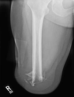
 The incidence of HO in combat-wounded service
originally described by one of the authors (BKP) has
members has consistently been reported as 63% – 64.6%,
been adopted. The severity of HO is graded using the
far greater than that described in civilian trauma centers.
The incidence of HO in combat-wounded service
originally described by one of the authors (BKP) has
members has consistently been reported as 63% – 64.6%,
been adopted. The severity of HO is graded using the
far greater than that described in civilian trauma centers.