Microsoft word - compcells_plasmidtransf.doc


Introduction
This week, we will treat bacteria to make them competent to take up plasmid
DNA. The process of DNA uptake is transformation, since it alters the genetic
compliment of the bacteria (they now have a new plasmid).
LEARNING GOALS:
1. Learn how to treat bacteria to take up plasmid DNA. 2. Understand the process of transformation. 3. Know how we select for bacteria that have taken up a plasmid.
Topic I: BACTERIAL COMPETENCE
1. Natural Competence
Bacteria are able to take up DNA from their environment (exogenous DNA) in
three ways; conjugation, transformation, and transduction. Only transformation is
the direct uptake of DNA, since conjugation requires cell-cell contact via a sex
pilus and transduction requires a bacteriophage intermediary to transfer DNA
from one cell to another.
For a bacterial cell to take up DNA from its surroundings, it must be in a special
physiological state called competence. Experiments by Frederich Griffith in 1929
using competent Streptococcus (now Enterococcus) pneumoniae were
instrumental in showing that DNA was the transforming principle – the genetic
material.
Natural competence is highly regulated in bacteria, and the factors leading to
competence vary among genera. For some genera, only a portion of the
population is competent at any time; for others, the entire population gains
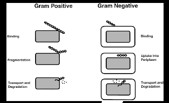
competence. A series of competence proteins is produced, which have some
homology but differ in the Gram negative and the Gram positive bacteria.
Figure 4.1: Transformation pathways in gram-positive and gram-negative
bacteria. From Dubnau, Ann Rev Microbiol, 1999.
Once the DNA has been brought into the cell's cytoplasm, it may be degraded by
cellular nucleases, or, if it is very similar to the cells own DNA, enzymes that
normally repair DNA may recombine it with the chromosome. Natural
transformation is very efficient for linear molecules such as fragments of
chromosomal DNA but not for circular plasmid DNAs.
2. Artificial Competence and Transformation
Artificial competence is not encoded in the cell's genes. Instead it is a laboratory
procedure in which cells are passively made permeable to DNA, using conditions
that do not normally occur in nature. These procedures are comparatively easy
and simple, and can be used to genetically engineer bacteria. However,
transformation efficiency is, in general, low. Only a portion of the cells become
competent and a fraction of those are successful in taking up DNA.
number of transformants (colonies) X final volume at recovery (mL) = number of transformants per microgram of plasmid DNA
Chilling cells in the presence of divalent cations such as CaCL2 or MgCL2
prepares the cell walls to become permeable to plasmid DNA. Cells are
incubated with the DNA. Then, during transformation, the cells are briefly heat
shocked
(42°C for 60-120 seconds), which causes the DNA to enter the cell.
This method works well for circular plasmid DNAs but not for linear molecules such as fragments of chromosomal DNA. Figure 4.2: Overview of competence and heat shock From: http://www.phschool.com/science/biology_place/labbench/lab6/test1.html
Cells that are undergoing very rapid growth are made competent more easily
than cells in other stages of growth. Thus cells are brought into log phase before
the procedure is begun. The log phase cells are all living, healthy, and actively
metabolizing. Because this procedure can be very harsh on cells, the log-phase
cells are more able to withstand this treatment.
The basic protocol for artificial competence has been known since the early
1970’s, and transformation efficiency has improved since those early days.
Competent cells are now readily available commercially. However, despite the
importance of this process to molecular biology and biotechnology, the exact
mechanisms involved in artificial competence are not known.
An article by Panja et al. (J Biotech. 2006) investigated the events producing
competence and transformation. In a previous work these researchers showed
that “naked” DNA is bound to the lipopolysaccharide (LPS) receptor molecules
on the competent cell surface. They suggested that the divalent cations formed
coordination complexes with the negatively charged DNA and LPS. However,
DNA is a large molecule and it was not known how the DNA crossed the cell
membrane to enter the cytosol. Their recent work shows that the heat-shock step
strongly depolarizes the cell membrane of CaCL2-treated cells. The decrease of
membrane potential may lower the negativity of the cell’s inside potential and
allow the movement of negatively charged DNA into the cell’s interior. A
subsequent cold-shock raises the membrane potential to its original value. The
authors were able to mimic the transformation process by using the protonophore
CCCP that reduces membrane potential by dissipating the proton-motive force
across the E. coli plasma membrane.
Topic II: IDENTIFICATION OF POSITIVE CLONES

1. Spreading a Plate
The technique used to obtain transformation colonies is to spread a plate rather
than streaking for single colonies. Both procedures will give isolated colonies that
are clonal (all bacteria in a colony are genetically identical). Spreading on a drug plate lets us use the entire plate surface to select for antibiotic-resistant
bacteria. In this experiment we also screen for the presence of a plasmid with an
insert. Transformation efficiency varies so we do not know how many bacteria
actually took up a plasmid, thus we usually spread several plates with different
volumes of cells. This is why it is important to thoroughly spread the cells over
the entire plate
until all of the solution is absorbed into the agar. Please examine
the photos on “how to spread a plate” available on the Virtual Lab Book.
2. Selection and Screening
Only bacteria that contain a viable plasmid with a drug resistance gene will be
able to grow on the drug plate. If the bacteria do not have a plasmid, or if the
plasmid is faulty (for example, no origin of replication), then we will never see it.
However, while we can select for bacteria containing a plasmid this does not
guarantee that the plasmid has our insert.
Even though we cannot select for plasmids containing a DNA insert, the plasmid
has the genetic tools to screen for potential inserts. The gene lacZ codes for the
-galactosidase enzyme, and this enzyme can metabolize the artificial substrate
X-gal (5-bromo-4-chloro-3-indolyl-b-D-galactopyranoside) to produce a blue
pigment. This is a straightforward concept, but the actual detection system has
an interesting twist. The lacZ DNA is in two separate locations; a small piece, the
 subunit, is located in the cloning vector. Within the  subunit is the Multiple
Cloning Site. The remaining lacZ DNA, the  subunit, is embedded in the
bacterial chromosome. The  and  subunits come together to make functional
-galactosidase that metabolizes the X-gal and produces blue colonies.
However, if a DNA insert disrupts the  subunit then NO functional -
galactosidase is produced and the colony is the normal “white” color. However,
there are reasons other than an insert that will cause a colony to be white,
including mutations in the  subunit or improper subunit assembly.
This discussion illustrates the important difference between a selection and a
screen. A well-designed selection will eliminate all unwanted possibilities. A
screen will help you eliminate many of the possibilities and guide you to the
desired outcome (a plasmid with an insert) but cannot guarantee that outcome.
REFERENCES
Dubnau D., DNA uptake in bacteria, Annu Rev Microbiol. 1999;53:217-44.
http://www.phschool.com/science/biology_place/labbench/lab6/test1.html
Panja S, Saha S, Jana B, Basu T., Role of membrane potential on artificial
transformation of E. coli with plasmid DNA, J Biotechnol. 2006 Dec 15;127(1):14-
20.

Source: http://delliss.people.cofc.edu/virtuallabbook/LabReadings/CompCellsTransf/CompCells_PlasmidTransf.pdf

septodont.at

Fachinformation Bezeichnung des Arzneimittels 2. Qualitative quantitative Zusammensetzung Die vollständige Auflistung der sonstigen Bestandteile siehe Abschnitt 6.1. Darreichungsform Sprühlösung zur Lokalanästhesie, Pumpspray 4. Klinische 4.1 Anwendungsgebiete - Oberflächliche Betäubung und Desinfektion der Mundschleimhaut vor einer - Oberflächliche Betäu

Microsoft word - 2009 equine progesterone instructions.doc

Quick Check™ Equine Progesterone Kit Recommended For Use in Veterinary Clinics For the determination of the mare’s estrus cycle The mare’s reproductive cycle is subject to great variability. Measuring the progesterone concentration permits the most accurate determination of where the mare is in her cycle and allows planning the most effective reproductive program. Knowing the proges

© 2010-2014 Pdf Medical Search