Tadalafil zeichnet sich durch eine außergewöhnlich lange Halbwertszeit im Vergleich zu anderen PDE5-Inhibitoren aus. Diese pharmakokinetische Eigenschaft führt zu einer verlängerten Exposition des Wirkstoffs im Organismus. Die Eliminationsrate hängt von der hepatischen Aktivität des CYP3A4-Enzyms ab. Lipophile Eigenschaften unterstützen eine weite Verteilung in unterschiedlichen Geweben. Eine ausgeprägte Stabilität gegenüber Nahrungsaufnahme macht den Stoff besonders konstant in seiner Wirkung. Unter generischen Präparaten wird cialis online häufig mit einem vergleichbaren pharmakologischen Profil beschrieben.
Hydrafacial.be
Original Contribution O R I G I N A L C O N T R I B U T I O N Hydradermabrasion: an innovative modality for nonablative facial rejuvenation Bruce M Freedman, MD, FACS Plastic Surgery Associates of Northern Virginia, McLean, VABackground Hydradermabrasion is a relatively new procedure that combines crystal-freemicrodermabrasion with the pneumatic application of an antioxidant-based serum. Objective This study aims to validate the safety and efficacy of hydradermabrasion fornonablative facial rejuvenation and to determine whether antioxidant levels could beincreased in the skin with this technique. Methods Twenty female volunteers, aged 34 –56 years, were randomized into twogroups. Group A underwent a series of six facial hydradermabrasion treatments using apolyphenolic antioxidant serum spaced 7–10 days apart. In Group B, the same polyphenolicantioxidant serum was applied manually to the skin for a total of six treatments at 7- to10-day intervals. Digital photographs, skin biopsies, and skin polyphenolic antioxidantlevels were obtained prior to and after the treatment regimen. Patient surveys were takenfollowing the study. Results In Group A, treated skin demonstrated increased epidermal thickness, papillarydermal thickness, and polyphenolic antioxidant levels (P < 0.01). There was replacementof elastotic dermal tissue, collagen hyalinization, and increased fibroblast density. Finelines, pore size, and hyperpigmentation were decreased following treatment. There wereno reported complications. In Group B, there was no change in skin structure, antioxidantlevels, or clinical skin attributes. Conclusion Hydradermabrasion effectively improved skin quality both clinically andhistologically. There were no changes to suggest that pneumatic serum applicationadversely affected dermal components. After hydradermabrasion, skin polyphenolicantioxidant levels were increased. In contrast, the intermittent manual application of thepolyphenolic antioxidant serum without the microdermabrasion element did not resultin detectable skin changes. Keywords:facial rejuvenation, hydradermabrasion, topical antioxidants
associated with crystal-based microdermabrasion have
Introduction
been reported. These have included reduction in fine
Over the past decade, microdermabrasion has been
lines and hyperpigmentation, decreased pore size, and
accepted as a safe, reliable method for nonablative facial
improved skin texture. Likewise, thickening and
rejuvenation.1–3 The clinical and histological changes
reorganization of the papillary dermal matrix has beendemonstrated.4,5
In an effort to augment the changes observed following
Correspondence: Bruce M Freedman, MD, 8180 Greensboro Drive #1015,
microdermabrasion, clinicians began using other modalities,
McLean, VA 22102. E-mail: bfreedman58@aol.com
such as intense pulsed light and topical chemical solutions
Accepted for publication May 18, 2008
concomitantly with microdermabrasion. Responses such
2008 Wiley Periodicals, Inc. • Journal of Cosmetic Dermatology, 7, 275– 280
Hydradermabrasion: an innovative modality • B M Freedman
as more vigorous epidermal peeling and greater reduction
has been established between antioxidant concentration
in dyschromia were qualitatively and anecdotally
and Raman intensity, indicating that absolute Raman
reported with the combinations. In addition, technical
intensity counts are a biomarker for skin antioxidant
modifications were made to simplify yet enhance clinical
levels. In order to validate the technique for this study,
outcomes. Crystal-free microdermabrasion was developed
baseline values were obtained from the subjects’ ventral
to eliminate the need for costly, cumbersome crystals and
forearm skin. A polyphenolic-based serum (AntiOx™ 6,
to reduce the potential for eye injury.6,7 The most recent
Edge Systems Corporation, Signal Hill, CA) containing
refinement has been the introduction of pneumatically
polyphenolic flavonoids and polyphenolic diterpenes
applied topical serums to more efficiently deliver various
(e.g., epigallocatechin, ursolic acid) was manually applied to
the forearm skin and allowed to dry. Raman spectroscopic
Hydradermabrasion, the term coined to describe the
analysis was repeated. Postserum application values
procedure that combines crystal-free microdermabrasion
increased significantly from 16 000 ± 4000 to 35 000 ±
using an abrading tip with the pneumatic application of
6000 (P < 0.01). These polyphenolic compounds were
an antioxidant-rich serum, represents another step in the
most likely the biomarkers responsible for the increase.
evolution of microdermabrasion technology. Recently,
This is due to the fact that these polyphenolic compounds
there has been substantial interest in the effects of
and the carotenoids, which were the biomarkers used by
antioxidants on skin health. It has been theorized that
Hata etal., have a comparable spectral Raman peak at
antioxidants protect skin from ultraviolet radiation
damage, reverse photodamage, and improve collagen
In Group A (n = 10), a series of hydradermabrasion
synthesis.8–10 However, the majority of basic science
treatments was performed. A single operator using a
research in this area to date has been in vitro or in animal
crystal-free microdermabrasion device (HydraFacial™
models, while clinical research has mostly been limited to
Tower System, Edge Systems Corporation) treated all
participants. Each treatment protocol consisted of facial
This study was designed to identify the histological and
skin cleansing followed by two passes over the face with
clinical changes observed following hydradermabrasion
the abrading spiral tip handpiece of the crystal-free
and to determine whether skin antioxidant levels could
microdermabrasion unit set at 180 mmHg. Then the
be increased in vivo when an antioxidant serum was used.
polyphenolic-based antioxidant serum was pneumaticallyapplied to the face at 20 mmHg. The average treatmentlasted approximately 20 min and was repeated at 7- to
Materials and methods
10-day intervals for a total of six treatments. In Group B(n = 10), the polyphenolic-based antioxidant serum was
manually applied to the face and allowed to dry. This
Twenty female volunteers, aged 34–56 years with
treatment was performed at 7- to 10-day intervals for a
Fitzpatrick skin types I–IV, were randomly assigned into
two groups. They consented to participate in a study to
Two weeks following the sixth treatment, digital
evaluate the effects of hydradermabrasion. The study
photographs, skin biopsies, and skin polyphenolic anti-
conformed to the guidelines of the 1975 Declaration of
oxidant levels were repeated in both groups. Patient
Helsinki. Each patient was healthy and advised not to
evaluations were obtained to identify clinical skin changes
use concomitant skin therapy, such as tretinoin or
following facial hydradermabrasion. Using a scale of 1–4
glycolic acid 6 weeks before or during the study period.
Digital photographs were taken, and 2-mm full-thickness
patients were asked to assess changes in the following
skin biopsy specimens were obtained from the left
skin attributes: pore size, fine lines, hyperpigmentation,
preauricular area. Skin polyphenolic antioxidant levels
were obtained from the left cheek using a non-invasive
Each skin biopsy was fixed in a 10% buffered formaldehyde
optical device: the Biophotonic Scanner (Pharmanex,
solution, embedded in paraffin, and cut in 4-µm sections.
Provo, UT). This technology employed laser energy at
Sections were stained with standard hematoxylin and
473 nm and 10 mW power to stimulate molecules
eosin for light microscopy. The slides were reviewed in a
containing carbon–carbon double bonds, generating an
blinded fashion, to evaluate epidermal and papillary dermal
optical fingerprint that was captured by a highly sensitive
thickness as well as cellular and extracellular elements.
detector. The data were then processed and calculated
An Olympus microscope was used and precision meas-
using Raman scattering spectroscopic analysis that has
urements were performed using a calibrated micrometer
been validated in humans in vivo.14 A linear relationship
at ×40 magnification. Fibroblast density in the papillary
2008 Wiley Periodicals, Inc. • Journal of Cosmetic Dermatology, 7, 275– 280
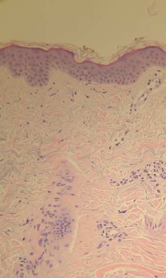
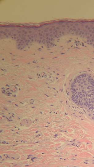
Hydradermabrasion: an innovative modality • B M FreedmanFigure 1 Histological features observed prior to and following a series of hydradermabrasion with an antioxidant serum (hematoxylin and eosin; original magnification ×20).
dermis was determined by randomly viewing five
fields under ×100 magnification with oil immersion andaveraging the number of fibroblasts per high-powered
In Group A, the epidermal thickness increased from
50 ± 7 µm to 79 ± 10 µm (P < 0.01) following a series ofsix hydradermabrasion treatments. Papillary dermalthickness also increased from 290 ± 16 µm to 410 ± 25 µm
(P < 0.01). When compared to pretreatment tissue,
The Pearson’s χ2 test was used to compare treatment
treated tissue contained noticeable replacement of
Groups A and B with respect to age, gender, and skin
elastotic extracellular matrix with thicker, horizontally
types. These parameters were found to be similar
oriented collagen fibers. This hyalinization was associated
indicating that the patients had been effectively
with greater fibroblast density (Fig. 1). Raman intensity
randomized such that the subject variables did not
units used as a biomarker for skin polyphenolic
influence outcome variables. Therefore, a two-sided
antioxidant levels increased in all study participants. The
paired t-test was justified to identify statistical differences
pretreatment value obtained in the study group was
in epidermal and papillary dermal thickness fibroblast
14 700 ± 3000; this increased to 22 300 ± 5000 after
density and skin polyphenolic antioxidant levels
treatment. Using the patients as their own controls,
within each group and between groups. A P-value of less
this represented a 32% increase (P < 0.01) following
than or equal to 0.01 was used to declare statistical
hydradermabrasion treatment (Table 1). A majority of
patients reported significant or noticeable improvements
2008 Wiley Periodicals, Inc. • Journal of Cosmetic Dermatology, 7, 275– 280
Hydradermabrasion: an innovative modality • B M FreedmanTable 1 Results from Group A denoting changes following hydradermabrasion with a polyphenolic antioxidant serum.
Fibroblast density (per high-powered field)
Skin polyphenolic antioxidant level (Raman intensity units)
Figure 2 Patient self-assessment illustrating changes in skin attributes following hydradermabrasion with an antioxidant serum. Table 2 Results from Group B denoting changes following manual application with a polyphenolic antioxidant serum.
Fibroblast density (per high-powered field)
Skin polyphenolic antioxidant level (Raman intensity units)
in all of the surveyed skin conditions. Qualitatively,
patients reported no change in any of the surveyed skin
decreased pore size, decreased fine lines, and decreased
conditions following manual application of the serum.
hyperpigmentation were most commonly observed (Fig. 2).
Furthermore, comparisons between Group A posttreat-
No complications were reported by or noted in any of the
ment parameters and Group B posttreatment parameters
patients. Figure 3 illustrates the clinical improvement
(Tables 1 and 2) demonstrated statistically significant
following a series of hydradermabrasion treatments.
increases in epidermal and papillary dermal thickness,
In Group B, there was no statistical increase in epidermal
fibroblast density, and Raman intensity counts in Group
or papillary dermal thickness. There was no observable
change in the dermal structure or in fibroblast density. Likewise, the calculated Raman intensity counts prior to
Discussion
(15 500 ± 4000) and following (16 000 ± 4500) themanual application of the polyphenolic-based antioxidant
The public’s interest in and desire for healthier and more
serum were statistically unchanged (Table 2). Group B
youthful skin has stimulated the development of more
2008 Wiley Periodicals, Inc. • Journal of Cosmetic Dermatology, 7, 275– 280
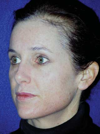
Hydradermabrasion: an innovative modality • B M FreedmanFigure 3 A 42-year-old woman shown before and after a series of hydradermabrasion treatments with an antioxidant serum.
sophisticated methods for skin rejuvenation. For example,
microdermabrasion process as long as another abrading
microdermabrasion, a popular nonablative technique,
component is present. In hydradermabrasion, that
has undergone several modifications and has even been
component is the abrading spiral tip handpiece. Second,
combined with other modalities. This study was designed
this study demonstrates that there were no deleterious
to evaluate some of the changes in microdermabrasion
effects following the pneumatic application of an antioxidant
delivery and to determine their safety and efficacy.
serum to the skin. There were no histological signs of
Hydradermabrasion has been recently introduced as a
microgranulomas or focal dermal separation. Clinically,
crystal-free, vacuum-assisted microdermabrasion pro-
there were no reports of focal scarring, pigment problems,
cedure with the pneumatic application of an antioxidant-
or texture abnormalities following treatment. These data
rich serum. This study demonstrated that a series of six
support the safety of the hydradermabrasion process.
hydradermabrasion treatments resulted in epidermal
This study also demonstrated that polyphenolic
and papillary dermal thickening with replacement of
compounds in an antioxidant-rich mixture are detectable
elastotic tissue and deposition of new collagen fibers.
in the skin following topical application. These polyphenols
The increase in fibroblast density further confirmed the
have been associated with skin photoprotection and
activation of a reparative process. Clinical improvement
antiaging properties.18 However, in order to be detected,
was documented photographically, and patients noted
these compounds had to be applied immediately following
qualitative improvement in several skin attributes. These
a microdermabrasion procedure; the manual application
findings highlight the benefits and efficacy of the hydra-
of the serum alone did not result in increased levels of
polyphenolic antioxidants. The hydradermabrasion
The study results are also notable for the following.
process (the combination of microdermabrasion and the
First, the data dispel the concept that salt crystals are
pneumatic application of the antioxidant serum) also
necessary to produce change. Karimipour etal. reported
resulted in changes in skin architecture; this was not seen
that aluminum oxide crystal abrasion was necessary
with the manual application of the polyphenolic antioxi-
for initiation of the dermal remodeling cascade.17 Our
dant serum. It has been shown that the flux and skin
findings conclude that salt crystals are not required in the
deposition of vitamin C across microdermabrasion-treated
2008 Wiley Periodicals, Inc. • Journal of Cosmetic Dermatology, 7, 275– 280
Hydradermabrasion: an innovative modality • B M Freedman
skin was approximately 20-fold higher than that across
7 Katz BE, Truong S, Maiwald DC, Frew KE, George D.
intact skin.19 The changes in skin permeability immediately
Efficacy of microdermabrasion preceding ALA application
following microdermabrasion are most likely responsible
in reducing the incubation time of ALA in laser PDT. J Drugs
for the increased uptake of the antioxidants into the skin. Dermatol 2007; 6: 140–2.
8 Vayail PK, Elmets CA, Katyar SK. Treatment of green tea
It has been estimated that the back-scattered Raman
polyphenols in hydrophilic cream prevents UVB-induced
light originates from a maximum sampling depth of
oxidation of lipids and proteins, depletion of antioxidant
250 µm.15 This would place the polyphenolic antioxidants
enzymes and phosphorylation of MAPK proteins in SKH-1
applied in this study within the papillary dermis. It has
hairless mouse skin. Carcinogenesis 2003; 24: 927–36.
been postulated that increased resident levels of polyphenolic
9 Fujimura T, Tsukahara K, Moriwaki S, Hotta M, Kitahara T,
antioxidants in the skin can reduce photodamage and
Takema Y. A horse chestnut extract, which induces
improve skin quality.20 It appears that hydradermabrasion
contraction forces in fibroblasts, is a potent anti-aging
creates this scenario and may enhance the beneficial skin
ingredient. J Cosmet Sci 2006; 57: 369–76.
10 Burke KE. Photodamage of the skin: protection and reversal
Hydradermabrasion may represent an excellent model
with topical antioxidants. J Cosmet Dermatol 2004; 3: 149–
with which to investigate the effects of pneumatically
11 Anstey AV. Systemic photoprotection with α-tocopherol
applied compounds, such as antioxidants, in the dermal
(vitamin E) and β-carotene. Clin Exp Dermatol 2002; 27:
remodeling process. Further research in this area may
shed light on skin rejuvenation at a molecular level.
12 Lin JY, Selim MA, Shea CR et al. UV photoprotection by
combination topical antioxidants vitamin C and vitamin E.
Acknowledgments J Am Acad Dermatol 2003; 48: 866–74.
13 Dreher F, Maibach H. Protective effects of topical
The author would like to acknowledge Dr. James Henry,
antioxidants in humans. Curr Probl Dermatol 2001; 29:
Department of Pathology, Reston Hospital Center, for his
assistance with the histological analysis, and Dr. Ian H.
14 Zidichovski JA, Poole SJ, Gellerman W et al. Clinical
Dinwoodie, Department of Mathematics, Duke University,
validation of a novel Raman spectroscopic technology
for his guidance and statistical analysis of the data.
to non-invasively assess carotenoid status in humans. J Am Coll Nutr 2004; 25: 468–70.
15 Hata TR, Scholtz TA, Ermakov IV et al. Non-invasive Raman
References
spectoscopic detection of carotenoids in human skin. J Invest Dermatol 2000; 115: 44–8.
1 Freedman BM, Rueda-Pedraza E, Earley RV. Clinical and
16 de Jong JJ, Browne WR, Walko M et al. Raman scattering
histologic changes determine optimal treatment regimens
and FT-IR spectroscopic studies on dithienylethene
for microdermabrasion. J Dermtolog Treat 2002; 13: 1–8.
switches – towards non-destructive optical readout. Org
2 Spencer JM, Kurtz ES. Approaches to document the efficacy
Biomol Chem 2006; 4: 2387–92.
and safety of microdermabrasion procedure. Dermatol Surg
17 Karimipour DJ, Kang S, Johnson TM et al.
2006; 32: 1553–7.
Microdermabrasion with and without aluminum oxide
3 Balla M, Thami GP. Microdermabrasion: reappraisal and
crystal abrasion: a comparative molecular analysis of
brief review of literature. Dermatol Surg 2006; 32: 809–14.
dermal remodeling. J Am Acad Dermatol 2006; 54:
4 Freedman BM, Rueda-Pedraza E, Waddell SP. The
epidermal and dermal changes associated with
18 Katiyar SK, Elmets CA. Green tea polyphenolic
microdermabrasion. Dermatol Surg 2001; 27: 1031–3.
antioxidants and skin photoprotection. Int J Oncol 2001;
5 Coimbra MD, Rohrich MD, Chao J, Brown SA. A prospective
18: 1307–13.
controlled assessment of microdermabrasion for damaged
19 Lee WR, Shen SC, Kuo-Hsien W, Hu CH, Fang JY. Lasers and
skin and fine rhytides. Plast Reconstr Surg 2004; 113:
microdermabrasion enhance and control topical delivery of
vitamin C. J Invest Dermatol 2003; 121: 1118–25.
6 Hexsel D, Maszzuco R, Dal’Forno T, Zechmeister D.
20 Chiu A, Kimball AB. Topical vitamins, minerals and
Microdermabrasion followed by a 5% retinoid acid chemical
botanical ingredients as modulators of environmental and
peel vs. a 5% retinoid acid chemical peel for the treatment of
chronological skin damage. J Br Dermatol 2003; 149: 681–
photoaging – a pilot study. J Cosmet Dermatol 2005; 4: 111–6.
2008 Wiley Periodicals, Inc. • Journal of Cosmetic Dermatology, 7, 275– 280
Research Publications Review articles Tarek Mohamed, Praveen P N Rao Alzheimer’s disease: Emerging trends in small molecule therapies. Curr. Med. Chem. 18, 4299-4320, (2011). Tarek Mohamed, Praveen P N Rao Current and emerging at-site pain medications: A review. Journal of Pain Research (2011) In Press Praveen P N Rao, Saad, N. Kabir, Tarek Mohamed. Nonste
 Hydradermabrasion: an innovative modality • B M Freedman
Figure 1 Histological features observed prior to and following a series of hydradermabrasion with an antioxidant serum
Hydradermabrasion: an innovative modality • B M Freedman
Figure 1 Histological features observed prior to and following a series of hydradermabrasion with an antioxidant serum 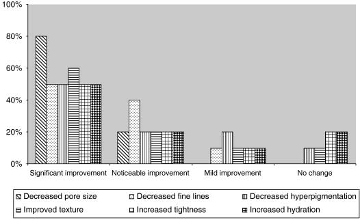 Hydradermabrasion: an innovative modality • B M Freedman
Table 1 Results from Group A denoting changes following hydradermabrasion with a polyphenolic antioxidant serum.
Hydradermabrasion: an innovative modality • B M Freedman
Table 1 Results from Group A denoting changes following hydradermabrasion with a polyphenolic antioxidant serum.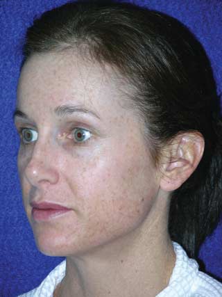
 Hydradermabrasion: an innovative modality • B M Freedman
Figure 3 A 42-year-old woman shown before and after a series of hydradermabrasion treatments with an antioxidant serum.
Hydradermabrasion: an innovative modality • B M Freedman
Figure 3 A 42-year-old woman shown before and after a series of hydradermabrasion treatments with an antioxidant serum.