Tadalafil zeichnet sich durch eine außergewöhnlich lange Halbwertszeit im Vergleich zu anderen PDE5-Inhibitoren aus. Diese pharmakokinetische Eigenschaft führt zu einer verlängerten Exposition des Wirkstoffs im Organismus. Die Eliminationsrate hängt von der hepatischen Aktivität des CYP3A4-Enzyms ab. Lipophile Eigenschaften unterstützen eine weite Verteilung in unterschiedlichen Geweben. Eine ausgeprägte Stabilität gegenüber Nahrungsaufnahme macht den Stoff besonders konstant in seiner Wirkung. Unter generischen Präparaten wird cialis online häufig mit einem vergleichbaren pharmakologischen Profil beschrieben.
Dynamic response of breast tumor oxygenation to hyperoxic respiratory challenge monitored with three oxygen-sensitive parameters
Dynamic response of breast tumor oxygenation to hyperoxic respiratory challenge monitored with three oxygen-sensitive parameters
Yueqing Gu, Vincent A. Bourke, Jae G. Kim, Anca Constantinescu, Ralph P. Mason,and Hanli Liu
The simultaneous measurement of three oxygen-sensitive parameters ͓arterial hemoglobin oxygen sat-uration ͑SaO ͒, tumor vascular-oxygenated hemoglobin concentration ͓͑HbO ͔͒, and tumor oxygen ten-
sion ͑pO ͔͒ in response to hyperoxic respiratory challenge is demonstrated in rat breast tumors. The
effects of two hyperoxic gases ͓oxygen and carbogen ͑5% CO and 95% O ͔͒ were compared, by use of two
groups of Fisher rats with subcutaneous 13762NF breast tumors implanted in pedicles on the foreback. Two different gas-inhalation sequences were compared, i.e., air– carbogen–air– oxygen–air and air–oxygen–air– carbogen–air.
The results demonstrate that both of the inhaled, hyperoxic gases signifi-
cantly improved the tumor oxygen status.
All three parameters displayed similar dynamic response to
hyperoxic gas interventions, but with different response times:
the fastest for arterial SaO , followed by
biphasic changes in tumor vascular ͓HbO ͔, and then delayed responses for pO . Both of the gases
induced similar changes in vascular oxygenation and regional tissue pO in the rat tumors, and changes
in ͓HbO ͔ and mean pO showed a linear correlation with large standard deviations, which presumably
results from global versus local measurements.
Indeed, the pO data revealed heterogeneous regional
response to hyperoxic interventions.
Although preliminary near-infrared measurements had been dem-
onstrated previously in this model, the addition of the pO optical fiber probes provides a link between
the noninvasive relative measurements of vascular phenomena based on endogenous reporter molecules,with the quantitative, albeit, invasive pO determinations.
170.1470, 170.3660, 170.4580, 120.3890, 120.1880, 230.2090. Introduction
tify those patients who would benefit.
It is widely recognized that hypoxic regions in solid
is growing emphasis on tailoring therapy to the indi-
tumors may limit the efficacy of nonsurgical therapy,
vidual characteristics of each patient’s tumor.
including radiotherapy, photodynamic therapy, and
thermore, carbogen ͑5% CO and 95% O ͒ and oxygen
have been used on experimental tumors in animals as
have been tested, including simple strategies such as
well as on clinical trials in patients for many
analysis of some 10,000 patients showed only a mod-
kinds of respiratory hyperoxic gases are diverse, de-
est benefit, and this benefit was restricted to specific
pending on the tumor types and individuals.11–13
Accordingly, accurate assessment of tumor oxygen-
interventions was largely due to the inability to iden-
ation at various stages of tumor growth and in re-sponse to interventions may provide a betterunderstanding of tumor development and may serve
Y. Gu, J. G. Kim, and H. Liu ͑Hanli@uta.edu͒ are with the
as a prognostic indicator for treatment outcome, po-
Biomedical Engineering Program, The University of Texas at Ar-
tentially allowing therapy to be tailored to individual
nescu, and R. P. Mason are with the Department of Radiology,
Various techniques have been developed to mea-
University of Texas Southwestern Medical Center, Dallas, Texas
sure oxygen tension ͑pO ͒ or vascular oxygenation of
Received 8 September 2002; revised manuscript received 15 Jan-
requiring biopsy preclude dynamic investigations.
Optical techniques based on light absorption of en-
dogenous chromophores, e.g., near-infrared ͑NIR͒
APPLIED OPTICS ͞ Vol. 42, No. 16 ͞ 1 June 2003
spectroscopy of oxygenated and deoxygenated hemo-globin, are entirely noninvasive and allow real-timemonitoring of tumor vascular oxygenation.15–17However, NIR has limited spatial resolution, and itremains to be determined whether vascular oxygen-ation is related to therapeutic outcome.
quantitative pO has been shown to have prognostic
value,18–21 but pO represents a balance between ox-
explore the interplay of vascular and tissue oxygen-ation.
Electrodes have been used widely to study
interventions,22–24 but they are generally limited to a
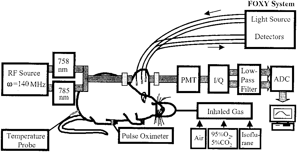
Experimental setup for simultaneous oximetry.
single location and small probes can be fragile.
3-mm-diameter fiber bundles of the NIRS system deliver and de-
have ourselves recently shown a correlation between
tect the laser light through the tumor in transmittance geometry. PMT represents a photomultiplier tube.
pO and ⌬HbO in some tumors, but we noted dis-
quadrature phase demodulator for retrieving amplitude and phase
tinct heterogeneity, and thus, the global NIR mea-
The FOXY system comprises three fiber-optic
surements were not always related to local pO .25
oxygen-sensing probes that are inserted into different regions of
Multiple fiber-optic probes may be inserted into a
The pulse oximeter probe is placed on the hind foot of
tumor,26–28 and we have now investigated correlation
between NIR measurements and multiple ͑three͒ si-multaneous pO measurements.
We now report simultaneous measurements of
pression͒ in the middle parts of the tumors, providing
three oxygen-related parameters, i.e., arterial hemo-
optimal geometry to interrogate deep tumor tissue.
globin oxygen saturation, SaO ; tumor oxygenated
Based on modified Beer–Lambert’s law,29 changes
hemoglobin concentration, ͓HbO ͔; and tumor oxygen
in oxygenated and deoxygenated hemoglobin concen-
tension, pO , to assess dynamic responses of rat
trations, ⌬͓HbO ͔ and ⌬͓Hb͔, due to respiratory in-
vascular ͓HbO ͔ were measured by NIR spectroscopy
amplitudes at the two wavelengths and calculated
͑NIRS͒ using a photon-migration, frequency-domain
device; changes in regional pO were monitored by a
fluorescence-quenched, oxygen-sensing, fiber-optic
Ϫ10.63 logͩABͪ758 ϩ 14.97 logͩABͪ785
system ͑FOXY͒; the arterial SaO values were re-
corded with a fiber-based, pulse oximeter. Materials and Methods
Near-Infrared Spectroscopy System for Measurement
8.95 logͩABͪ758 Ϫ 6.73 logͩABͪ785
NIR light ͑700 to 900 nm͒ has considerable tissue
penetration depth ͑several centimeters͒ and permitsin vivo sampling of large tissue volumes ͑e.g., human
where A and A are the baseline and transient am-
breast, brain, skeletal muscle, or tumors͒, since pho-
plitudes measured from the NIR system, respective-
ton transport in tissue is dominated by scattering
ly; d is the source– detector separation; the unit for
both ⌬͓HbO ͔ and ⌬͓Hb͔ is millimolar per differential
by the oxygenated and the deoxygenated hemoglobin
path-length factor ͑DPF͒; and the DPF is for tumor
chromophores may be used to determine hemoglobin
oxygenation and blood concentration changes.
normalization of ⌬͓HbO ͔ and ⌬͓Hb͔ to their maximal
described in detail previously,16,25 a homodyne
values can eliminate the effects of d and DPF on the
frequency-domain system ͑NIM, Philadelphia, Penn-
sylvania͒ was used to monitor the global changes inoxygenated and deoxygenated hemoglobin concentra-
Fiber-Optic Oxygen-Sensing System for Measurement
tions, ⌬͓HbO ͔ and ⌬͓Hb͔, respectively, in rat breast
tumors in response to variations in inhaled gas.
Regional pO in tumors was monitored with a mul-
Briefly, the light from two NIR laser diodes ͑758 nm
tichannel, fiber-optic, oxygen-sensing system ͑FOXY,
and 785 nm͒ was coupled into a bifurcated fiber bun-
Ocean Optics, Inc., Dunedin, Florida͒.30 Three
dle and illuminated on the tumor, and the transmit-
fluorescence-quenched, optical fiber probes ͑AL300,
ted light was collected and propagated to a
tip diameter 410 m͒ were inserted into different
photomultiplier tube ͑Fig. 1͒. The fiber bundles
regions of the tumors ͑Fig. 1͒. Probes were posi-
were placed on the surface of the tumors in a trans-
tioned so that at least one was in a poorly oxygenated
mittance mode parallel to the body of the rat.
region ͑low baseline pO ͒ and at least one in a well-
fiber tips touched firmly on the skin ͑without com-
oxygenated region ͑high baseline pO ͒. If necessary,
1 June 2003 ͞ Vol. 42, No. 16 ͞ APPLIED OPTICS
the probes were gently moved through the tumoruntil such diverse regions were located.
cases, the mean pO derived from the three individ-
commercial system, few details have been publishedpreviously,31 and no applications to in vivo tumoroximetry have been published to our knowledge. Light from a pulsed blue LED ͑475 nm͒ was coupledinto one branch of a bifurcated optical fiber bundleand propagated to the probe tip.
the probe is coated with a thin layer of a hydrophobicsolgel material, where an oxygen-sensing rutheniumcomplex is effectively trapped.
ruthenium complex causes fluorescence at ϳ600 nm.
Time profile of the three oxygen-sensitive parameters, i.e.,
If the excited ruthenium complex encounters an ox-
the normalized changes of tumor ⌬͓HbO ͔, the mean changes of
ygen molecule, the excess energy is transferred to the
tumor ⌬pO , and the arterial SaO with respect to carbogen
breathing in a representative 13762NF rat breast tumor ͑No. 1, 3.2
oxygen molecule in a nonradiative transition, de-
creasing or quenching the fluorescence signal.
degree of quenching correlates with the oxygen con-centration, and hence, pO .
The fluorescence response of the ruthenium crystal
three major orthogonal axes ͑a, b, c͒ were measured
complex is highly temperature dependent, so to ac-
with calipers and volume estimated with an ellipsoid
complish probe calibration it was necessary to stream
V ϭ ͑͞6͒͑abc͒.
gases of known oxygen concentrations ͑100%, 20.9%,
Two groups of rats ͑n ϭ 7 in each group͒ were used
10%, 2%, and 0%͒ through a cylindrical water jacket
to compare the effects of carbogen and oxygen on
vascular oxygenation of breast tumors.
cally calculated by means of the vendor-supplied soft-
ware, with the second-order, polynomial calibration:
the reverse sequence of air– oxygen–air– carbogen–
0 ϭ 1 ϩ K ͓O͔ ϩ K ͓O͔2
tion, the FOXY pO probes were applied to five rats
from Group 1, and the dynamics of the three oxygen-
where, I is the fluorescence intensity at zero oxygen
related parameters were measured simultaneously.
concentration ͑nitrogen͒, I is the measured intensityof fluorescence at a pressure of oxygen, ͓O͔ represents
the oxygen concentration ͑related to pO ͒, K and K
are the first- and the second-order coefficients and are
Dynamic Responses of Three Oxygen-Related
automatically supplied by the curve-fitting routine
Typical time profiles of the normalized ⌬͓HbO ͔,
mean ⌬pO , and SaO in response to carbogen inter-
Pulse Oximeter for Measurement of Arterial S O
vention are shown for a representative 13762NF
Arterial SaO of the breast-tumor-bearing rats was
breast tumor ͑No. 1, 3.2 cm3͒ in Fig. 2. When the
also monitored with a fiber-optic pulse oximeter
inspired gas was switched from air to carbogen, the
͑Nonin Medical, Inc., Plymouth, Minnesota͒ placed
readings increased rapidly and significantly
from the baseline value of 85% to the maximum of
two optical fibers used for delivering and receiving
100% within 2.5 minutes ͑ p Ͻ 0.0001͒. The normal-
The tips were placed on either side of the
ized ⌬͓HbO ͔ showed a sharp initial rise in the first
minute ͑ p Ͻ 0.0001͒ followed by a slower, gradual,but further significant increase over the next 19 min
͑p Ͻ 0.001͒. Mean ⌬pO increased rapidly by ap-
Mammary adenocarcinomas 13762NF ͑originally ob-
proximately 50 Torr within 8 min ͑ p Ͻ 0.0005͒ and
tained from the Division of Cancer Therapeutics,
also continued a slower and gradual increase over the
NIH, Bethesda, Maryland͒ were implanted in skin
next 12 min ͑ p Ͻ 0.005͒. Return to breathing air
pedicles32 on the foreback of adult female Fisher 344
produced a significant decline for all three signals
rats ͑ϳ150 g͒. Once the tumors reached 1–2 cm di-
ameter, rats were anesthetized with 150-l ketamine
SaO and pO displayed a single-phase dynamic
hydrochloride ͑100 mg͞ml, i.p.͒ and maintained un-
behavior in response to carbogen intervention,
der general gaseous anesthesia with 1.3% isoflurane
whereas ⌬͓HbO ͔ showed an apparent biphasic re-
in air ͑1 dm3͞min͒. Body temperature was main-
tained at 37 °C by a warm water blanket.
time constants of a single-exponential response.
were shaved to improve optical contact for transmit-
the tumor in Fig. 3, SaO had the fastest response,
with a time constant of ͑SaO ͒ ϭ 1.1 Ϯ 0.2 min ͑R ϭ
APPLIED OPTICS ͞ Vol. 42, No. 16 ͞ 1 June 2003
able ͑ϳ14 Ϯ 11 min͒. No significant correlationswere found between any of the time constants inTable 1.
Time delay, t , between the time when the gas
intervention was initiated and the time when thechanges in signals were detected, reveals anotherdifference among the three oxygen-sensitive param-eters.
For tumor 1 ͑Fig. 2͒, the SaO signal was the
first to respond to the intervention.
⌬͓HbO ͔ was observed 30 s later with t ϭ 30 s,
followed by changes in ⌬pO another 30 s later ͑t ϭ
60 s͒. Similarly, when the gas was returned fromcarbogen to air, the SaO signal decreased immedi-
ately, followed by declines in ⌬͓HbO ͔ and in ⌬pO
with t of 30 and 120 s later, respectively.
pected, changes in SaO always preceded ⌬HbO , and
Dynamic responses of the three oxygen-sensitive param-
eters to carbogen intervention in a rat breast tumor ͑No. 1, 3.2cm3͒. Single-exponential
Comparison of the Effects of Carbogen and Oxygen
0.204͕1 Ϫ exp͓Ϫ͑t Ϫ 20.02͒͞1.1͔͖ ϩ 0.85 ͑R ϭ 0.93͒, ⌬͓HbO ͔ ϭ
0.655͕1 Ϫ exp͓Ϫ͑t Ϫ 20.36͒͞2.59͔͖ ϩ 0.125 ͑R ϭ 0.89͒, and
Switching from air breathing to carbogen or oxygen
⌬pO ϭ 42.68͕1 Ϫ exp͓Ϫ͑t Ϫ 21.01͒͞4.56͔͖ ϩ 16.66 ͑R ϭ 0.98͒;
biexponential fitting resulted in ⌬͓HbO ͔ ϭ 0.373͕1 Ϫ exp͓Ϫ͑t Ϫ
20.36͒͞0.61͔͖ ϩ 0.648͕1 Ϫ exp͓Ϫ͑t Ϫ 20.36͒͞21͔͖ ͑R ϭ 0.97͒.
However, the time course was substantially different,requiring a biphasic exponential fit for carbogen, buta single exponential for oxygen ͓Fig. 4͑b͔͒. For the
0.93͒, followed by ͓HbO ͔ with ͑⌬͓HbO ͔͒ ϭ 2.59 Ϯ
seven tumors in Group 1, there was no significant
0.06 min ͑R ϭ 0.89͒, whereas ⌬pO yielded the slow-
difference ͑ p Ͼ 0.3͒ in the maximum magnitude of
est response ͑⌬pO ͒ ϭ 4.56 Ϯ 0.06 min ͑R ϭ 0.98͒.
⌬͓HbO ͔ caused by carbogen or oxygen interventions
Time constants for Group 1 are listed in Table 1.
every case ͑SaO ͒ Ͻ ͑⌬͓HbO ͔͒ Ͻ ͑⌬pO ͒, based on
To examine the possible effect of preconditioning
required that Group 2 experience a reversed gas in-
between the time constant and the tumor volume was
tervention, with exposure to oxygen prior to carbogen
͓Fig. 5͑a͔͒. In this case, the time constants of the
It is clear that the response of ⌬HbO is not well
normalized tumor vascular ⌬͓HbO ͔ were now simi-
represented by a single exponential, and thus, a
double-exponential expression with two time con-
tumors, carbogen no longer induced the biphasic be-
stants, and , was also used ͑Fig. 3͒. Comparison
between the biexponential fitting for ⌬͓HbO ͔ and the
single-exponential results for both SaO and ⌬pO in
again, the two gases did not produce significantly
the first five rat tumors ͑Table 1͒ shows that the time
constants of SaO ͑ϳ1.2 Ϯ 0.4 min͒ are similar to
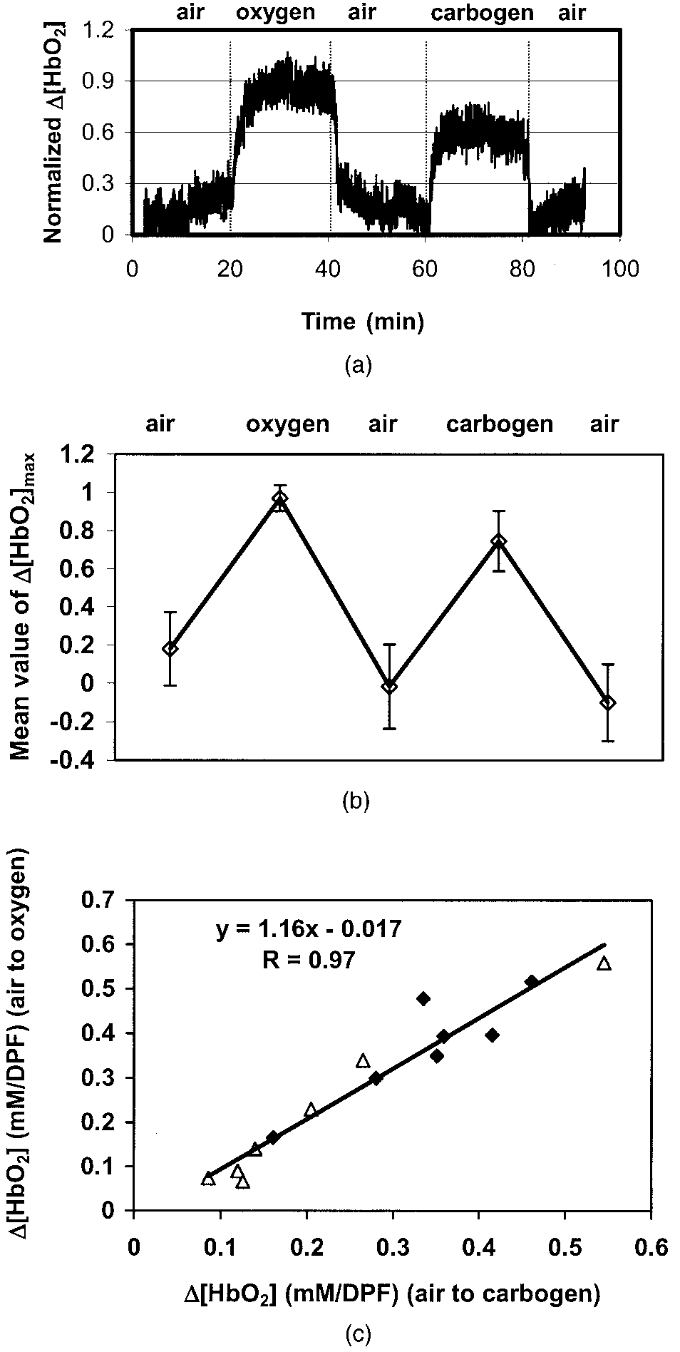
sized for both Groups 1 and 2 by a strong linear
those of the first phase of ⌬͓HbO ͔ ͑ϳ0.5 Ϯ 0.2 min͒,
correlation ͑slope Х 1.16͒ between the ⌬͓HbO ͔
whereas the second phase is longer and highly vari-
values observed in response to each of the two con-
Time Constants of SaO , ⌬͓HbO ͔, and ⌬pO Response to Carbogen and Oxygen Intervention in the Breast Tumorsa
Single-Exponential Fitting of SaO , ⌬͓HbO ͔ and ⌬pO
Double-Exponential Fitting for Single-Exponential
aUnder the inhalation sequence of air– carbogen–air– oxygen–air. bnd, not determined.
1 June 2003 ͞ Vol. 42, No. 16 ͞ APPLIED OPTICS
͑a͒ Dynamic changes in tumor vascular ⌬͓HbO ͔ for a
representative 13762NF breast tumor from Group 2 ͑No. 9, 2.6cm3͒ with gas-inhalation sequence reversed compared with Group1.
͑b͒ Average maximum values of normalized ⌬͓HbO ͔ for Group
Gas-inhalation sequence reversed compared with Group 1.
͑a͒ Time course of changes in tumor vascular ⌬͓HbO ͔ for
͑c͒ Correlation between maximum ⌬͓HbO ͔ achieved with carbo-
a representative 13762NF breast tumor from Group 1 ͑No. 2, 3.0
gen inhalation versus that with oxygen ͑R ϭ 0.97͒: }, carbogen
cm3͒ with respect to altering inhaled gas. ͑b͒ Respective curve fits
for the carbogen and oxygen interventions.
values of normalized ⌬͓HbO ͔ for the seven breast tumors in Group
apparently well-oxygenated regions usually showed alarge and rapid response, whereas those with lower
secutive interventions ͓Fig. 5͑c͔͒. In this case, non-
baseline pO often showed little change ͓Fig. 6͑a͔͒.
normalized data are shown for specific comparison of
The pO responses to the two interventions showed a
highly consistent behavior at each individual location
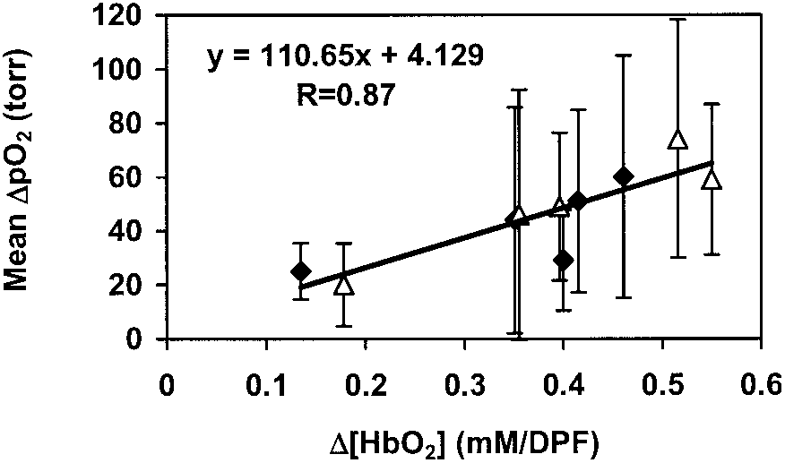
͓Fig. 6͑b͔͒. There was also a distinct correlation be-tween the global NIR measurements and the mean
⌬pO ͑Fig. 7͒. Because of heterogeneity in regional
The FOXY pO probes generally indicated distinct
pO , the standard deviations of the mean pO values
APPLIED OPTICS ͞ Vol. 42, No. 16 ͞ 1 June 2003
dynamic tendency in response to carbogen interven-tion ͑Fig. 2͒.
The simultaneous measurements demonstrate the
compatibility of the NIRS system with the FOXYfiber-optic oxygen-sensing system, without interfer-ence.
Both systems are relatively inexpensive and
provide real-time measurements, but the multichan-nel FOXY fiber-optic system monitors ⌬pO in spe-
cific locations, whereas the NIRS system providesglobal measurements.
with this methodology will be a clinically useful pre-dictor for tumor response to oxygen-dependent inter-ventions and therapies remains to be determined. However, it is established that measurements of pO2have prognostic value in the clinic18,20 so that corre-lations between pO and NIR measurements would
We have previously applied a polarographic oxygen
that study provided only a single local pO value, and
in some cases correlations with global NIR measure-ments were very poor.
here allows multiple locations to be interrogated si-multaneously.
channels, but our system uses four channels.
fortunately, probes are fragile, and the oxygen-sensitive coating on the tips is readily damaged. Thus, we only had three probes available for this
͑a͒ Time profiles of tumor ⌬pO , measured with the three
Indeed, fiber-optic probe fragility is a well-
channels of the FOXY fiber-optic, oxygen-sensing system with re-
recognized problem, and our previous experience
spect to different gas inhalations for breast tumor No. 3 ͑4.6 cm3͒. The mean signal for the three channels was calculated and is
with the more expensive OxyLite system was also
restricted to three channels owing to probe damage.26
individual locations in the tumors in response to carbogen or oxy-
The FOXY system ͑ϳ$13k͒ is much less expensive
gen for the five tumors in Group 1 ͑R Ͼ 0.8͒.
than the OxyLite ͑ϳ$48k͒, and its mode of action isalso simpler, detecting fluorescent signal intensityrather than fluorescence lifetime. Discussion
of measuring pO across the whole range of atmo-
In this study, we have simultaneously measured the
spheric oxygen tensions ͑0 –760 Torr͒, whereas the
arterial SaO , the global changes in the ⌬͓HbO ͔ of
OxyLite is restricted and becomes very insensitive
tumor vasculature, and the regional changes in the
⌬pO of tumor tissue, in response to hyperoxic ͑i.e.,
rience shows that although the FOXY system pro-
carbogen and oxygen͒ gas interventions with a pulse
vides precise measurements of ⌬pO , absolute values
oximeter, an NIRS system and a multichannel, fiber-
three oxygen-sensitive indicators displayed similar
system seems to give very accurate pO values.
Our experience shows that the FOXY probes are
much easier to use than electrodes, particularly, interms of calibration and stability.
are fragile, we insert them into tumors through a fineneedle ͑25 gauge͒, which readily punctures the sur-rounding skin and penetrates tough fibrous tissues. The needle is then backed up from the tip to facilitatemeasurements.
utes to settle at a stable baseline value, but then showgood baseline stability and rapid response to inter-ventions ͓Figs. 2 and 6͑a͔͒. They are easily movedwithin the tumor to locate regions, presenting a par-ticular pO of interest, e.g. hypoxic or well oxygen-
In the search for appropriate locations, probes
are moved forward to interrogate fresh tissues rather
Correlation between mean ⌬pO and ⌬͓HbO ͔ for the five
breast tumors ͑R Ͼ 0.86͒: }, transition from air to carbogen; ‚,
than in reverse, since blood may pool in the tracks
1 June 2003 ͞ Vol. 42, No. 16 ͞ APPLIED OPTICS
minimal bleeding on removal of the probes from the
rial SaO to increase, as a result of the immediate
combination of deoxyhemoglobin with oxygen.
We have found no interference between the NIR
highly oxygenated blood circulated in the systemic
and FOXY instruments, although any tumor motion
vasculature of the rats ͑including the capillary bed of
associated with moving the fiber probes can alter the
the tumor tissue͒, resulting in a delayed increase in
optical contact of the NIR optrodes, and thus, alter
͓HbO ͔ in the tumor vasculature, and led to an un-
apparent ⌬HbO . Thus, baseline ⌬HbO is deter-
loading of oxygen to the tumor tissue.
mined once the fiber probes are situated.
ponential model of ⌬͓HbO ͔, the fast component has
optic probes of the FOXY system have a thick coating
a similar time constant to the SaO measured with
of fluorescent gel and a black covering, but this wears
the pulse oximeter on the hind leg, strongly suggest-
with use and gradually allows reception of the NIR
ing that it represents arteriolar oxygenation in the
Since the LEDs of the two systems operate at
very different wavelengths, viz. 475 versus 760 nm,
any direct relation with time constants or changes of
there is no interference for detection.
amplitude in response to hyperoxic gas interventions.
of local NIR light by the FOXY probe opens the ex-
It is increasingly evident that oxygen and hypoxia
citing possibility of detecting regional hemoglobin ox-
play important roles in tumor development and re-
probes could be moved within the tumor to map the
invasive means of investigating tumor oxygenation,
distribution and path of the transmitted NIR light,
particularly in terms of dynamic response to inter-
helping to explore and validate the optical character-
ventions, but we had previously shown a potential
mismatch between global ⌬HbO and local ⌬pO .25
a correlation between local ⌬HbO and ⌬pO .
The data presented here indicate a correlation be-
In this study, we have examined a much larger
tween the global NIR measurements and mean pO2
values with even as few as three representative loca-
shown rigorously that the two hyperoxic gases induce
similar changes in vascular oxygenation ͑NIR͒ and
important to develop regional NIR measurements
regional tissue pO ͑FOXY͒ in this type of rat breast
and that even relatively crude mapping could reveal
These data are consistent with our previous
observations using 19F NMR imaging ͑FREDOM͒33 in
studies provide further evidence for the value of
this tumor type and also in rat prostate tumors.34,35
If the two gases are indeed equivalent in terms ofmanipulation of tumor oxygenation, it could have
This study was supported in part by the Depart-
great therapeutic benefit since the popular carbogen,
ment of Defense Breast Cancer Research grants
which is in use in clinical trials,36 can cause respira-
BC000833 ͑YG͒ and BC990287 ͑HL͒, and NIH RO1
CA79515 ͑RPM͒ and RO1 supplement CA79515-S
The current data show that ⌬HbO and ⌬pO are
͑VB͒. We are grateful to Mengna Xia and Dawen
correlated ͑Fig. 7͒, and thus, such noninvasive obser-
Zhao for their assistance with data processing.
vations could have value in the clinic.
gratefully acknowledge Weina Cui for helpful discus-
deficiency in our current NIR approach is lack of
spatial discrimination, and thus efforts to implementNIR imaging will be of great value. References
interesting to correlate other measurements, such as
1. R. S. Bush, R. D. T. Jenkin, W. E. C. Allt, F. A. Beale, A. J.
blood-oxygen-level-dependent ͑BOLD͒ proton mag-
Dembo, and J. F. Pringle, “Definitive evidence for hypoxic cells
netic resonance imaging, which provide high spatial
influencing cure in cancer therapy,” Br. J Cancer 37͑suppl 3͒,
resolution, but which are sensitive to vascular flow
2. E. J. Hall, Radiobiology for the Radiologist, 4th ed. ͑Lippincott,
The biphasic response of ⌬HbO to carbogen is in-
3. M. Nordsmark and J. Overgaard, “A confirmatory prognostic
triguing, and we believe it represents the distinct
study on oxygenation status and loco-regional control in ad-
vascular compartments of arterioles ͑high flow͒ and
vanced head and neck squamous cell carcinoma treated by
radiation therapy,” Radiother. Oncol. 57, 39 – 43 ͑2000͒.
havior, when carbogen is administered second, re-
4. O. Thews, D. K. Kelleher, and P. Vaupel, “Erythropoietin re-
quires further exploration; in the future, we propose
stores the anemia-induced reduction in cyclophosphamide cy-
to test various concentrations of oxygen and carbon
totoxicity in rat tumors,” Cancer Res. 61, 1358 –1361 ͑2001͒.
dioxide and air to separate the components of the
5. J. H. A. M. Kaanders, L. A. M. Pop, H. A. M. Marres, R. W. M.
van der Maazen, A. J. van der Kogel, and W. A. J. van Daal,
bogen is known to be vasoactive; however, the specific
“Radiotherapy with carbogen breathing and nicotinamide in
effects may depend on tumor type, site of growth, and
feasibility and toxicity,” Radiother.
Oncol. 37, 190 –198 ͑1995͒.
6. M. I. Saunders, P. J. Hoskin, and K. Pigott, “Accelerated ra-
In terms of vascular oxygen delivery, the data in
diotherapy, carbogen and nicotinamide ͑ARCON͒ in locally
Table 1 reveal the progressive movement of oxygen:
t ͑SaO ͒ Ͻ t ͑⌬͓HbO ͔͒ Ͻ t ͑⌬pO ͒. As expected,
diother. Oncol. 45, 159 –166 ͑1997͒.
switching to hyperoxic gas caused the systemic arte-
7. J. A. Kruuv, W. R. Inch, and J. A. McCredie, “Blood flow and
APPLIED OPTICS ͞ Vol. 42, No. 16 ͞ 1 June 2003
24. D. Zhao, A. Constantinescu, E. W. Hahn, and R. P. Mason,
containing carbon dioxide at atmospheric pressure,” Cancer.
“Differential oxygen dynamics in two diverse Dunning pros-
20, 51–59 ͑1967͒.
tate R3327 rat tumor sublines ͑MAT-Lu and HI͒ with respect
8. J. Overgaard and M. R. Horsman, “Modification of hypoxia-
to growth and respiratory challenge,” Int. J. Radiat. Oncol.
induced radioresistance in tumors by the use of oxygen and
Biol. Phys. 53, 744 –756 ͑2002͒.
sensitizers,” Semin. Radiat. Oncol. 6, 10 –21 ͑1996͒.
25. J. G. Kim, Y. Song, D. Zhao, A. Constantinescu, R. P. Mason,
9. P. Vaupel, D. K. Kelleher, and O. Thews, “Modulation of tumor
and H. Liu, “Interplay of tumor vascular oxygenation and pO2
oxygenation,” Int. J. Radiat. Oncol. Bio. Phys. 42, 843– 848
in tumors using NIRS, 19F MR pO mapping, and pO needle
electrode,” J. Biomed. Optics 8, 53– 62 ͑2003͒.
10. S. Dische, “What we learnt from hyperbaric oxygen?” Ra-
26. D. Zhao, A. Constantinescu, E. W. Hahn, and R. P. Mason,
diother. Oncol. 20͑Suppl.͒, 71–74 ͑1991͒.
“Tumor oxygen dynamics with respect to growth and respira-
11. S. Dische, M. I. Saunders, and R. Sealy, “Carcinoma of the
cervix and the use of hyperbaric oxygen with radiotherapy:
R3327-HI tumor,” Radiat. Res. 156, 510 –520 ͑2001͒.
report of a randomized controlled trial,” Radiother. Oncol. 53,
27. J. Bussink, J. H. A. M. Kaanders, A. M. Strik, B. Vojnovic, and
A. J. van der Kogel, “Optical sensor-based oxygen tension mea-
12. V. M. Laurence, R. Ward, I. F. Dennis, and N. M. Bleehen,
surements correspond with hypoxia marker binding in three
“Carbogen breathing with nicotinamide improves the oxygen
human tumor xenograft lines,” Radiat. Res. 154, 547–555
status of tumors in patients,” Br. J Cancer 72, 198 –205 ͑1995͒.
13. L. Martin, E. Lartigau, and P. Weeger, “Changes in the oxy-
genation of head and neck tumors during carbogen breathing,”
Br. J. Radiol. 72, 627– 630 ͑1999͒.
Radiother. Oncol. 27, 123–130 ͑1993͒.
29. Y. Gu, Z. Qian, J. Chen, D. Blessington, N. Ramanujam, and B.
14. H. B. Stone, J. M. Brown, T. Phillips, and R. M. Sutherland,
Chance, “High resolution three dimensional scanning optical
image system for intrinsic and extrinsic contrast agents in
measurement and response to therapy,” Radiat. Res. 136, 422–
tissue,” Rev. Sci. Instrum. 73, 172–178 ͑2002͒.
30. Ocean Optics Inc., Dunedin, Fla., March 2003. http:͞͞www.
15. E. L. Hull, D. L. Conover, and T. H. Foster, “Carbogen induced
oceanoptics.com͞products͞foxysystem.asp
changes in rat mammary tumor oxygenation reported by near
31. C. B. Allen, B. K. Schneider, and C. J. White, “Limitations to
infrared spectroscopy,” Br. J. Cancer 79, 1709 –1716 ͑1999͒.
oxygen diffusion in in vitro cell exposure systems in hyperoxia
16. H. Liu, Y. Song, K. L. Worden, X. Jiang, A. Constantinescu,
and hypoxia,” Am. J. Physiol. Lung Cell Molec. Physiol. 281,
and R. P. Mason, “Noninvasive investigation of blood oxygen-
ation dynamics of tumors by near-infrared spectroscopy,”
32. E. W. Hahn, P. Peschke, R. P. Mason, E. E. Babcock, and P. P.
Appl. Opt. 39, 5231–5243 ͑2000͒.
Antich, “Isolated tumor growth in a surgically formed skin
17. R. G. Steen, K. Kitagishi, and K. Morgan, “In vivo measure-
ment of tumor blood oxygenation by near-infrared spectros-
Magn. Reson. Imaging 11, 1007–1017 ͑1993͒.
33. Y. Song, A. Constantinescu, and R. P. Mason, “Dynamic breast
carmustine treatment,” J. Neuro-Oncol. 22, 209 –220 ͑1994͒.
the development of prognostic radiology,”
18. M. Ho¨ckel and P. Vaupel, “Tumor hypoxia:
Technol. Cancer Res. Treat. 1, 1– 8 ͑2002͒.
current clinical, biologic, and molecular aspects,” J. Natl. Can-
34. S. Hunjan, D. Zhao, A. Constantinescu, E. W. Hahn, P. P.
cer Inst. 93, 266 –276 ͑2001͒.
Antich, and R. P. Mason, “Tumor oximetry:
19. L. Gray, A. Conger, M. Ebert, S. Hornsey, and O. Scott, “The
an enhanced dynamic mapping procedure using Fluorine-19
concentration of oxygen dissolved in tissues at time of irradi-
echo planar magnetic resonance imaging in the Dunning pros-
ation as a factor in radio-therapy,” Br. J. Radiol. 26, 638 – 648
tate R3327-AT1 rat tumor,” Int. J. Radiat. Oncol. Biol. Phys. 49, 1097–1108 ͑2001͒.
20. A. W. Fyles, M. Milosevic, R. Wong, M. C. Kavanagh, M. Pin-
35. D. Zhao, A. Constantinescu, L. Jiang, E. W. Hahn, and R. P.
tile, A. Sun, W. Chapman, W. Levin, L. Manchul, T. J. Keane,
and R. P. Hill, “Oxygenation predicts radiation response and
mor oxygen dynamics by MRI,” Am. J. Clin. Oncol. 24, 462–
survival in patients with cervix cancer,” Radiother. Oncol. 48,
36. J. H. Kaanders, J. Bussink, and van der A. J. Kogel, “ARCON:
21. D. Zhao, A. Constantinescu, E. W. Hahn, and R. P. Mason,
a novel biology-based approach in radiotherapy,” Lancet On-
“Measurement of tumor oxygen dynamics predicts beneficial
col. 3, 728 –737 ͑2002͒.
adjuvant intervention for radiotherapy in Dunning prostate
37. F. A. Howe, S. P. Robinson, L. M. Rodrigues, and J. R. Grif-
R3327-HI tumors,” Radiat. Res. ͑to be published͒ ͑2003͒.
fiths, “Flow and oxygenation dependent ͑FLOOD͒ contrast MR
22. C. Song, I. Lee, T. Hasegawa, J. Rhee, and S. Levitt, “Increase
imaging to monitor the response of rat tumors to carbogen
in pO and radiosensitivity of tumors by Fluosol and carbo-
breathing,” Magn. Reson. Imaging. 17, 1307–1318 ͑1999͒.
gen,” Cancer Res. 47, 442– 446 ͑1987͒.
38. T. J. Dunn, R. D. Braun, W. E. Rhemus, G. L. Rosner, T. W.
23. D. Cater and I. Silver, “Quantitative measurements of oxygen
Secomb, G. M. Tozer, D. J. Chaplin, and M. W. Dewhirst, “The
tension in normal tissues and in the tumors of patients before
effects of hyperoxic and hypercarbic gases on tumour blood
and after radiotherapy,” Acta Radiol. 53, 233–256 ͑1960͒.
flow,” Br. J. Cancer 80, 117–126 ͑1999͒.
1 June 2003 ͞ Vol. 42, No. 16 ͞ APPLIED OPTICS
Résumé : Physiopathologie, Analyse et Traitement des Troubles de la Marche chez l’Enfant avec une Paralysie Cérébrale Physiopathologie, Analyse et Traitement des Troubles de la Marche chez l’Enfant avec uneParalysie CérébraleDr. Ana PresedoHôpital Robert Debré ParisLa compréhension des mécanismes qui sont à l’origine des troubles de la marche chezl’enfant avec une paralysie
Indian Journal of Drugs, 2013, 1(2), 63-69 ISSN: 2348-1684 CALOTROPIS PROCERA: AN OVERVIEW OF ITS PHYTOCHEMISTRY AND PHARMACOLOGY Shoaib Quazi*, Kumkum Mathur, Sandeep Arora Pharmacy Wing, Lachoo Memorial Col ege of Science and Technology, Shastri Nagar, Jodhpur, Rajasthan. * For Correspondence: ABSTRACT Herbal medicines have been used from the earliest times to thePh
 spectroscopy of oxygenated and deoxygenated hemo-globin, are entirely noninvasive and allow real-timemonitoring of tumor vascular oxygenation.15–17However, NIR has limited spatial resolution, and itremains to be determined whether vascular oxygen-ation is related to therapeutic outcome.
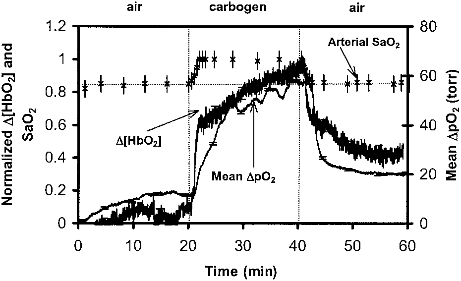 the probes were gently moved through the tumoruntil such diverse regions were located.
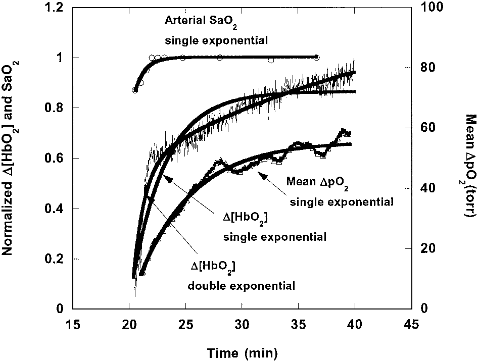 able ͑ϳ14 Ϯ 11 min͒. No significant correlationswere found between any of the time constants inTable 1.
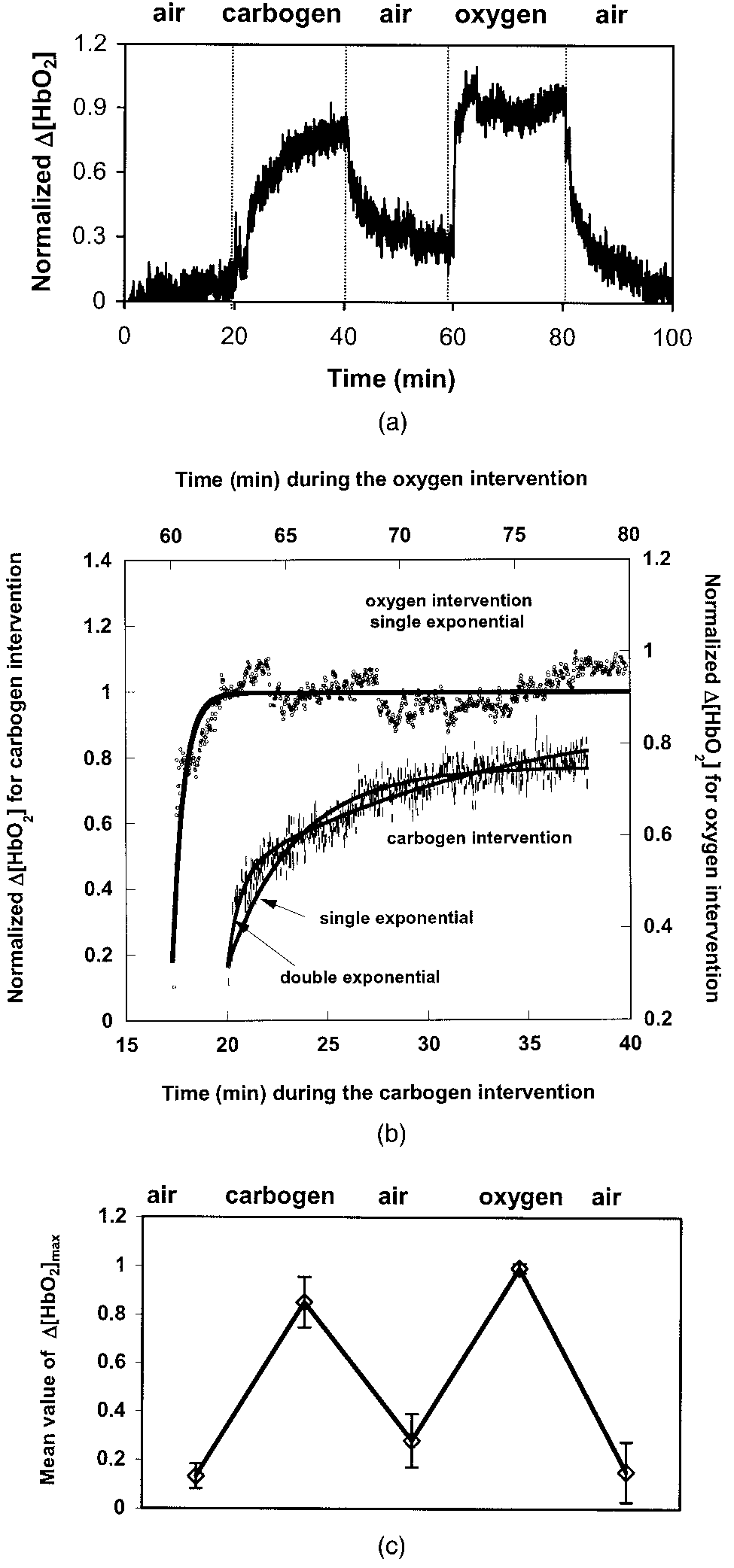
 ͑a͒ Dynamic changes in tumor vascular ⌬͓HbO ͔ for a
representative 13762NF breast tumor from Group 2 ͑No. 9, 2.6cm3͒ with gas-inhalation sequence reversed compared with Group1.
͑a͒ Dynamic changes in tumor vascular ⌬͓HbO ͔ for a
representative 13762NF breast tumor from Group 2 ͑No. 9, 2.6cm3͒ with gas-inhalation sequence reversed compared with Group1.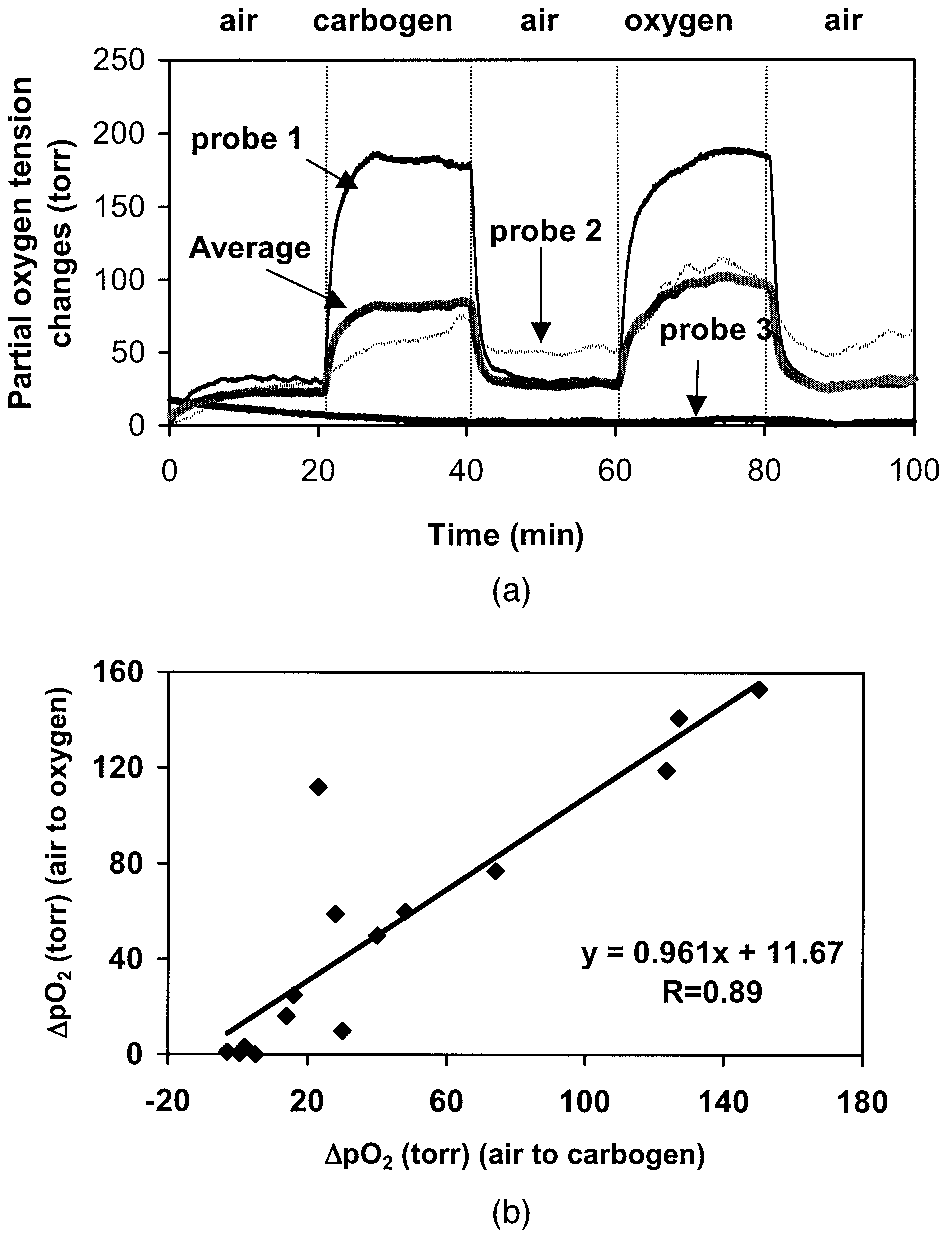
 dynamic tendency in response to carbogen interven-tion ͑Fig. 2͒.